
Adele
RESPONDING TO THE ENVIRONMENT - PLANTS GRADE 12 NOTES - LIFE SCIENCES STUDY GUIDES
RESPONDING TO THE ENVIRONMENT - PLANTS
LIFE SCIENCES
STUDY GUIDES AND NOTES
GRADE 12
- Growth and development in plants
- Role of auxins in phototropism and geotropism
- Plant defence mechanisms
CHAPTER 9: RESPONDING TO THE ENVIRONMENT - PLANTS
9.1 Growth and development in plants
Growth and development in plants are controlled by hormones. Auxin is an example of a hormone.
Tropism is the growth or turning movement of a plant or part of a plant in response to an environmental stimulus.
- Phototropism is the growth of a plant in the direction of a light source.
- Geotropism is the growth of a plant in response to gravity.
The growth movement of phototropism and geotropism is due to chemical messengers (hormones) called auxins in a plant.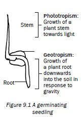
Activity 1
Questions
Complete the table:
Term | Description |
a) | Chemical messenger in the plant |
b) | Growth of a plant stem towards light |
Geotropism | c) |
Tropism | d) |
Answers to activity 1
a) Plant hormone✔
b) Phototropism✔
c) Growth of a plant root in response to gravity✔
d) Growth movement of a part of a plant in response to an environmental stimulus✔ [4]
9.2 Role of auxins in phototropism and geotropism
Role of auxins in phototropism | Role of auxins in geotropism |
Produced at the tip of stem/shoot | Produced at the tip of roots |
Auxins move downward evenly | Auxins move upwards evenly |
This even distribution brings about equal growth on all sides of the stem | This even distribution brings about equal growth on all sides of the root |
As a result the stem grows upward | As a result the root grows downward |
When the stem is exposed to unilateral light (light from one side only) | When the root is placed horizontally (only one side exposed to gravity) |
The auxin concentration will be high on the dark side - light destroys auxins | The auxin concentration will be high on the lower side of the root - gravity attracts auxins |
More growth occurs on the dark side because auxins stimulate growth on the dark side | More growth occurs on the upper side of the root because auxins on the lower side inhibit growth |
As a result the stem bends towards the light | As a result the root bends downwards |
Activity 2
Questions
QUESTIONS 1 to 2 are based on Diagrams I and II which illustrate the response of the tip of a young shoot to a light stimulus.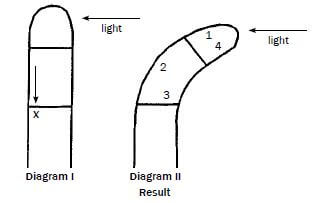
- The arrow X represents the unequal distribution of …
- abscisic acid.
- mineral salts.
- gibberellins.
- auxins.
- The curving of the shoot in Diagram II is due to more rapid cell growth in region(s) …
- 1.
- 2.
- 1 and 4.
- 3 and 4.
- The diagram above represents …
- geotropism.
- apical dominance.
- phototropism.
- gravity. 3 × 2 [6]
Answers to activity 2
1. D✔✔
2. B✔✔
3. C✔✔ 3 × 2 [6]
9.3 Plant defence mechanisms
Plants are eaten by herbivores and attacked by pathogenic organisms such as viruses, bacteria and fungi, causing them to become diseased.
Plants protect themselves from these threats using chemicals and thorns.
Activity 3
1. Describe TWO methods used by plants as defence mechanisms (4)
Answers to activity 3
1.
- Plants have thorns✔ on their stems and leaves. They are unable to escape from herbivores, and the thorns are used to protect themselves. ✔
- The chemical secretion of plants is poisinous to some organisms. ✔
- Sticky secretions given of by plants make it difficult for insects and animals to eat the plant ✔ [4]
HOMEOSTASIS IN HUMANS GRADE 12 NOTES - LIFE SCIENCES STUDY GUIDES
HOMEOSTASIS IN HUMANS
LIFE SCIENCES
STUDY GUIDES AND NOTES
GRADE 12
CHAPTER 8: HOMEOSTASIS IN HUMANS
8.1 Introduction
Homeostasis is the process of maintaining a constant internal environment within the body. The internal environment refers to the blood and tissue fluid that surrounds the cells of the body. Homeostasis enables the body to function efficiently, despite changes that might occur in the external or internal environment.
Changes in temperature, glucose levels, carbon dioxide levels, water levels and salt levels of the internal environment affects the homeostatic balance of the body. Negative feedback mechanisms operate in the human body to detect changes or imbalances in the internal environment and to restore the balance.
8.2 Negative feedback mechanisms
General sequence of events in a negative feedback mechanism :
Step 1: An imbalance is detected.
Step 2: A control centre is stimulated.
Step 3: Control centre responds.
Step 4: Message sent to target organs/s.
Step 5: The target organ responds.
Step 6: It opposes/reverses the imbalance.
Step 7: Balance is restored.
8.2.1 The regulation of glucose levels in the internal environment
When the glucose level in the blood increases above normal levels: | |
Step 1 | Glucose levels in the blood increase above normal levels |
Step 2 | The pancreas is stimulated |
Step 3 | to secrete insulin into the blood |
Step 4 | Insulin travels in the blood to the liver |
Step 5 | where it stimulates the conversion of excess glucose to glycogen which is then stored |
Step 6 | The glucose level in the blood now decreases |
Step 7 | and returns to normal |
When the glucose level in the blood decreases below normal levels: | |
Step 1 | Glucose levels in the blood decrease below normal levels |
Step 2 | The pancreas is stimulated |
Step 3 | to secrete glucagon into the blood |
Step 4 | Glucagon travels in the blood to the liver |
Step 5 | where it stimulates the conversion of stored glycogen to glucose |
Step 6 | The glucose level in the blood now increases |
Step 7 | and returns to normal |
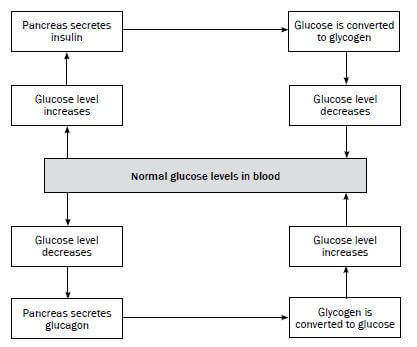
Figure 8.1 Negative feedback mechanism to regulate the glucose levels
8.2.2 The regulation of carbon dioxide levels in the internal environment
When the CO2 level in the blood increases above normal levels: | |
Step 1 | CO2 levels in the blood increase above normal levels |
Step 2 | Receptor cells in the carotid artery in the neck are stimulated |
Step 3 | To send impulses to the medulla oblongata in the brain |
Step 4 | Medulla oblongata stimulates breathing muscles (intercostal muscles and diaphragm) and heart |
Step 5 | Breathing muscles contract more actively - increases the rate and depth of breathing. The heart beats faster. |
Step 6 | More CO2 is taken to and exhaled from the lungs |
Step 7 | The CO2 level in the blood returns to normal |
8.2.3 The regulation of water balance in the internal environment (osmoregulation)
When the blood has less water than normal: | |
Step 1 | Blood has less water than normal |
Step 2 | The hypothalamus is stimulated |
Step 3 | and sends impulses to the pituitary gland to secrete more ADH |
Step 4 | ADH travels in the blood to the kidneys |
Step 5 | ADH increases the permeability of the collecting ducts and the distal convoluted tubules of the kidney |
Step 6 | More water is re-absorbed and passed to the surrounding blood vessels |
Step 7 | The water level in the blood returns to normal |
When the blood has more water than normal: | |
Step 1 | Blood has more water than normal |
Step 2 | The hypothalamus is stimulated |
Step 3 | and sends impulses to the pituitary gland to stop secreting ADH/to secrete less ADH |
Step 4 | No ADH/less ADH travels in the blood to the kidneys |
Step 5 | The collecting ducts and the distal convoluted tubules of the kidney become less permeable to water |
Step 6 | Less water is re-absorbed and passed to the surrounding blood vessels. More water is now lost |
Step 7 | The water level in the blood returns to normal |
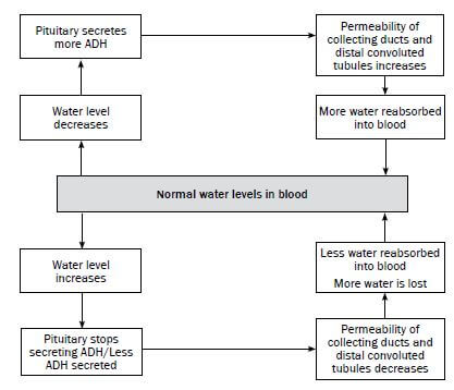
Figure 8.2 Negative feedback mechanism to regulate the water balance
8.2.4 The regulation of salt balance in the internal environment
When the salt level in the blood decreases: | |
Step 1 | The salt level in the blood decreases |
Step 2 | Receptor cells in the afferent and efferent arterioles of the kidney detect the low salt level |
Step 3 | The adrenal gland is stimulated |
Step 4 | into secreting more aldosterone |
Step 5 | Aldosterone increases the re-absorption of sodium ions from the renal tubules in the kidney into the surrounding blood vessels |
Step 6 | The salt level in the blood vessels increases |
Step 7 | and returns to normal |
When the salt level in the blood increases: | |
Step 1 | The salt level in the blood increases |
Step 2 | Receptor cells in the afferent and efferent arterioles of the kidney detect the high salt level |
Step 3 | The adrenal gland is stimulated |
Step 4 | to stop secreting aldosterone/to secrete less aldosterone |
Step 5 | This decreases the re-absorption of sodium ions from the renal tubules in the kidney into the surrounding blood vessels |
Step 6 | The salt level in the blood vessels decreases |
Step 7 | and returns to normal |
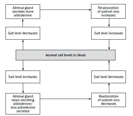
Figure 8.3 Negative feedback mechanism to regulate the salt balance
8.3 The process of temperature regulation
Temperature regulation is the control of body temperature to keep it as close to 37ºC as possible to enable the body to function normally.
Body temperature is regulated by the hypothalamus in the brain and the blood vessels and sweat glands in the skin.
Figure 8.4 below shows how the body temperature is regulated by the hypothalamus and the skin.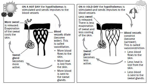
Figure 8.4 The homeostatic mechanism to regulate body temperature
Activity 1
Questions
1. Name the heat regulation centre in the brain. (1)
2. What happens to the blood vessels of the skin on a cold day? (1)
3. Describe how the state of the blood vessels mentioned in question 2 decreases heat loss. (4)
4. What happens to blood vessels of the skin on a hot day? (1)
5. Describe how the state of the blood vessels mentioned in question 4 increases heat loss. (4) [11]
Answers to activity 1
1. Hypothalamus✔ (1)
2. Blood vessels constrict✔/vasoconstriction (1)
3.
- Less blood flows to the surface of the skin.✔
- Less heat is lost from the surface of the skin.✔
- Less blood flows to the sweat glands.✔
- Sweat glands release less sweat.✔
- Less evaporation of sweat.✔
- Less cooling of the skin on a cold day.✔ (any 4)(4)
4. Blood vessels dilate✔/vasodilation (1)
5.
- More blood flows to the surface of the skin.✔
- More heat is lost from the surface of the skin.✔
- More blood flows to the sweat glands.✔
- Sweat glands release more sweat.✔
- Evaporation of sweat✔
- cools the skin on a hot day.✔ (any 4)(4) [11]
ENDOCRINE SYSTEM GRADE 12 NOTES - LIFE SCIENCES STUDY GUIDES
ENDOCRINE SYSTEM
LIFE SCIENCES
STUDY GUIDES AND NOTES
GRADE 12
CHAPTER 7: ENDOCRINE SYSTEM
7.1 The human endocrine system
The endocrine system is responsible for chemical coordination and regulates activities that take place inside the body. The endocrine system consists of glands
that produce different hormones, which are the body’s chemical messengers. Figure 7.1 below shows the glands of the endocrine system, the hormones they produce and the function of these hormones in the body.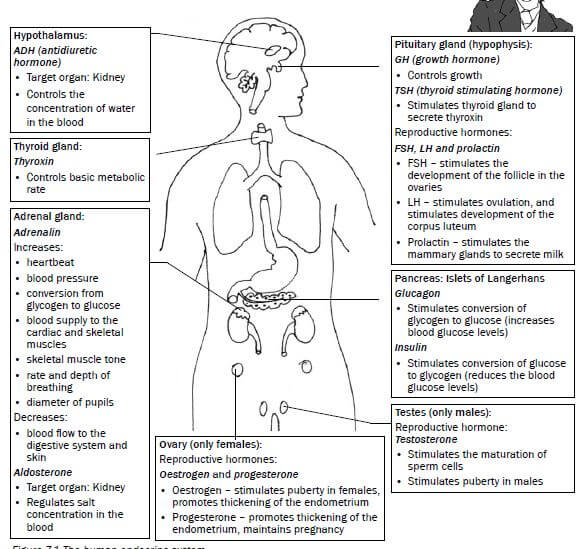
Figure 7.1 The human endocrine system
7.2 Negative feedback
Homeostasis is a process of maintaining a constant internal environment (blood and tissue fluid) within the body. This enables the body to function efficiently, despite changes in the external or internal environment.
Negative feedback mechanisms operate in the human body to detect changes or imbalances in the internal environment and to restore the balance.
7.2.1 General sequence of events in a negative feedback mechanism
Step 1: An imbalance is detected.
Step 2: A control centre is stimulated.
Step 3: Control centre responds.
Step 4: Message sent to target organ/s.
Step 5: The target organ responds.
Step 6: It opposes/reverses the imbalance.
Step 7: Balance is restored.
7.2.2 Example of a negative feedback mechanism
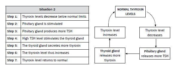
We will look at the regulation of thyroxin in the human body. There are two glands involved in the control of thyroxin levels:
- Gland 1: Thyroid gland (releases thyroxin)
- Gland 2: Pituitary gland (releases TSH)
Let us now look at the sequence of events in this feedback mechanism.
When you read the flow diagrams, start with NORMAL THYROXIN LEVELS.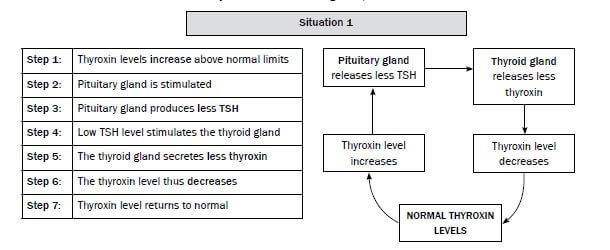
Activity 1
Question
The flow chart in Figure 7.2 below shows the control of glucose levels.
Provide labels for 1 to 6. [6]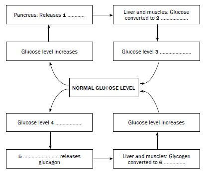
Figure 7.2 The negative feedback system to control glucose levels in the body
Answers to activity 1
- Insulin✔
- Glycogen ✔
- Decreases✔
- Decreases✔
- Pancreas ✔
- Glucose✔ [6]
RESPONDING TO THE ENVIRONMENT- HUMANS GRADE 12 NOTES - LIFE SCIENCES STUDY GUIDES
RESPONDING TO THE ENVIRONMENT- HUMANS
LIFE SCIENCES
STUDY GUIDES AND NOTES
GRADE 12
CHAPTER 6: RESPONDING TO THE ENVIRONMENT - HUMANS
The human nervous system
The nervous system is responsible for processing and transmitting information throughout the body:
- It tells the body how to react to stimuli (changes in the environment to which the body responds). For example, it regulates body temperature on a hot or cold day. It is also responsible for the reflex action, for example, when you step on a pin or touch a hot surface.
- The nervous system also coordinates the various activities of the body, such as walking, hearing, seeing, and so on.
The central nervous system consists of the brain and the spinal cord.
6.1 The brain
6.1.1 Structure and functions of the brain
Figure 6.1 below shows the different parts of the brain and their functions.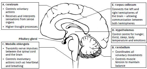 Figure 6.1 The structure and functions of the brain
Activity 1
Questions
Write down the name of the part which:
- Controls heartbeat (1)
- Contains the centres that control balance, muscle tone and equilibrium (1)
- Has centres that interpret what you see (1)
- Coordinates voluntary muscle movements (1)
- Controls body temperature (1) [5]
Answers to activity 1
- Medulla oblongata✔(1)
- Cerebellum✔ (1)
- Cerebrum✔ (1)
- Cerebellum✔ (1)
- Hypothalamus✔ (1) [5]
6.2 Neurons
Neurons are specialised cells which connect the brain and spinal cord to all other parts of the body.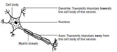 Figure 6.2 A neuron
There are three types of neurons, namely sensory (afferent) neurons, motor (efferent) neurons and interneurons (or connectors). Table 6.1 below shows the structure and function of these neurons.
| Type of neuron | Function | Structure |
| Sensory (afferent) neuron Senses the stimulus | Transmits impulses from the sense organs or receptors to the spinal cord and brain. | 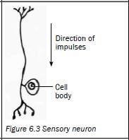 |
| Motor (efferent) neuron Response to the stimulus | Transmits impulses from the brain and spinal cord to the effectors (muscles and glands). The effectors bring about the response. | 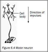 |
| Interneuron (connector) Found in the brain and the spinal cord. | Links the sensory neuron to the motor neuron | 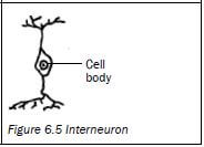 |
Table 6.1 Sensory, motor and interneurons
A synapse is the functional connection between the axon of one neuron, and the dendrites of another neuron.
6.3 Reflex arc
A reflex action is a quick, automatic action that involves the spinal cord and does not involve the brain. It is an important function to protect the body from harm. Examples are blinking the eye, coughing, sneezing, dilation and constriction of the pupil of the eye, and quickly withdrawing your hand when it touches a hot surface.
The reflex arc is the path along which an impulse is transmitted to bring about a response to a stimulus during a reflex action.
Figure 6.6 below shows what happens when you hold your finger close to a flame. The grey arrows represent the reflex arc.
The path of a reflex arc: |
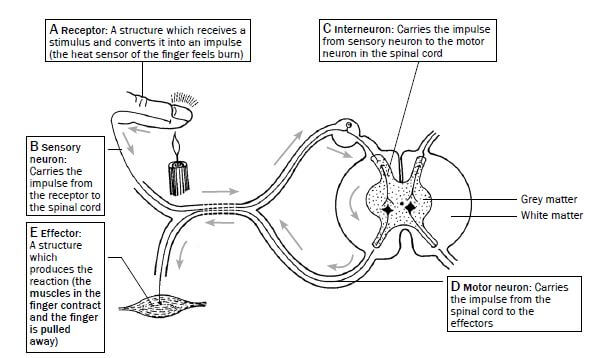
Figure 6.6 The reflex action of withdrawing a finger when placed in a flame
Activity 2
Questions
Use the diagram of the reflex arc in Figure 6.6 on page 44 to answer the following questions.
- Part B indicates the …
- dendrite of the motor neuron.
- axon of the motor neuron.
- dendrite of the sensory neuron.
- axon of the sensory neuron. (2)
- The correct sequence in which impulses move from the receptor to the effector in the reflex arc in Figure 6.6 is ...
- A → B → C → D→ E
- C → A → B → E→ D
- C → B → E → D→ A
- A → D → E → B→ C (2)
- Give the correct term for the following definitions:
- A structure which receives a stimulus and converts it into a impulse
- A structure which responds to a stimulus, e.g. a muscle or gland
- A neuron that carries impulses from the central nervous system to the effectors
- A neuron that carries impulses from the receptors to the central nervous system
- A neuron that carries impulses from a sensory neuron to a motor neuron in the spinal cord
- A very quick, automatic action that involves the spinal cord and not the brain
- The pathway along which an impulse is transmitted to bring about a response to a stimulus during a reflex action 7 × 1 = (7) [11]
Answers to activity 2
- C✔✔(2)
- A✔✔ (2)
-
- Receptor✔
- Effector✔
- Motor/efferent neuron✔
- Sensory/afferent neuron✔
- Interneuron✔/ connector
- Reflex action✔
- Reflex arc✔ 7 × 1 = (7) [11]
6.4 The human eye
Figure 6.7 below shows the different parts of the eye and their functions.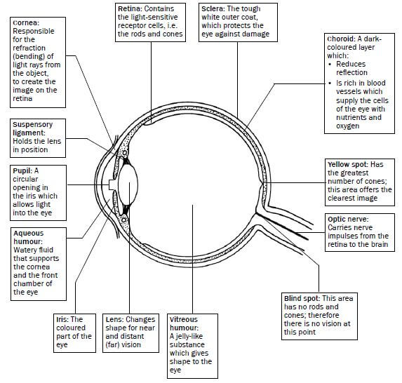
Figure 6.7 The structure of the eye
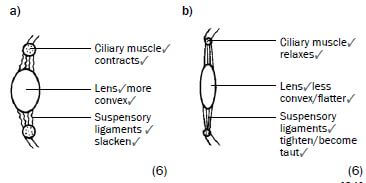
6.4.1 Accommodation
Accommodation is the adjustment of the shape of the lens to see objects clearly whether they are far away or close by. This is shown in Table 6.2 and Figures 6.8 and 6.9 below.
Distant vision (objects further than 6 m) | Near vision (objects closer than 6 m) |
1. Ciliary muscles relax | 1. Ciliary muscles contract |
2. Suspensory ligaments tighten (become taut) | 2. Suspensory ligaments slacken |
3. Tension on lens increases | 3. Tension on lens decreases |
4. Lens is less convex (flatter) | 4. Lens becomes more convex (more rounded) |
5. Light rays are refracted (bent) less | 5. Light rays are refracted (bent) more |
6. Light rays are focused onto the retina | 6. Light rays are focused onto the retina |
|
|
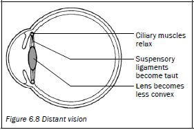
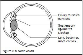
Table 6.2 Accommodation of the eye for distant and near vision
6.4.2 Pupillary mechanism
The pupillary mechanism (or pupil reflex) regulates the amount of light entering the eye by adjusting the size of the pupil. This is shown in Table 6.3 and Figures 6.10 and 6.11 below.
Light is bright | Light is dim |
1. Radial muscles of the iris relax | 1. Radial muscles of the iris contract |
2. Circular muscles of the iris contract | 2. Circular muscles of the iris relax |
3. Pupil constricts (gets smaller) | 3. Pupil widens (gets bigger) |
4. Less light enters the eye | 4. More light enters the eye |
|
|
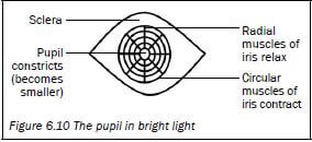
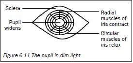
Table 6.3 Pupillary mechanism
6.4.3 Visual defects
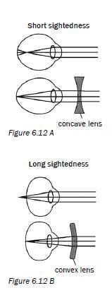 | Visual defect | Nature of the defect | Corrective measures |
| Short-sightedness Near objects can be seen clearly (myopia) |
| Wearing glasses with converging (biconcave) lensFigure 6.12 A | |
| Long-sightedness Objects far-away can be seen clearly (hyperopia) |
| Wearing glasses with converging (biconvex) lensFigure 6.12 B | |
| Astigmatism |
| Glasses with lenses shaped to correct the distortion | |
| Cataracts |
| Surgery to replace thelens with a synthetic lens |
Activity 3
Questions
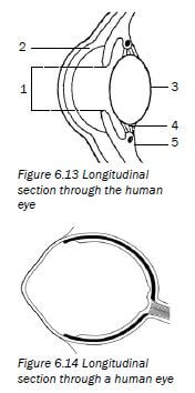
- Figure 6.13 shows a longitudinal section through the human eye. Study the diagram and answer the questions that follow.
a) Label parts 2, 3, 4 and 5 respectively. (4)
b) Name and describe the process that causes part 1 to dilate (become wider). (5) - Figure 6.14 is a longitudinal section through the human eye. The structures which enable the eye to focus on objects are missing in this diagram. Study the diagram and answer the questions that follow.
Draw a longitudinal section through the missing parts of Figure 6.14 to indicate the appearance of these structures when you are ...
a) reading a book. (6)
b) looking at an object more than 6 metres away. (6) [21]
Answers to activity 3
-
a) 2 - Cornea✔
3 - Lens✔
4 - Suspensory ligaments✔
5 - Ciliary muscles✔/ body (4)
b) Pupillary mechanism✔/ pupil reflex
The radial muscles3of the iris contract3and the circular muscles✔relax.✔
The pupil dilates and more light enters the eye.✔ (5) -
 [21]
[21]
 [21]
[21]6.5 The human ear
6.5.1 Structure of the ear
The human ear consists of three main parts:
- The outer ear
- The middle ear
- The inner ear
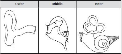
Figure 6.15 below shows the structure and function of each part of the human ear.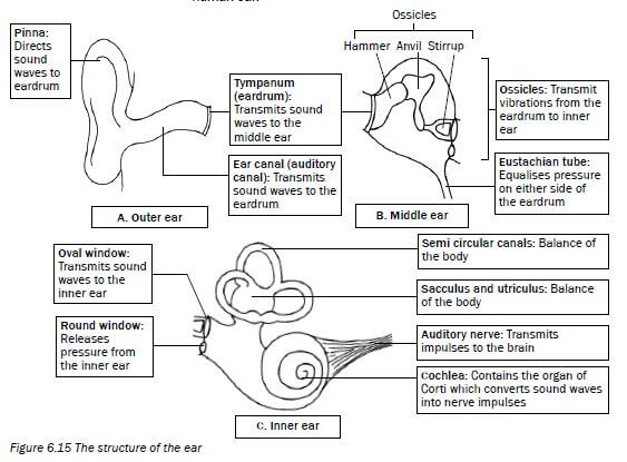
Figure 6.15 The structure of the ear
6.5.2 Hearing
Figure 6.16 below shows how the three parts of the ear work together to make it possible for us to hear. The grey arrows show the path of a sound wave.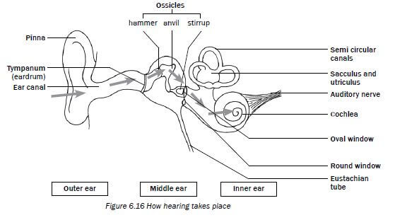
Figure 6.16 How hearing takes plac
Look at Figure 6.16 above and read the information in Table 6.4 below to understand how hearing takes place.
Part of ear | What it does during the hearing process |
Pinna | Traps the sound waves and directs them into the auditory canal. |
Tympanic membrane | Vibrates and transmits the vibrations to the ossicles in the middle ear. |
Ossicles | The ossicles amplify the vibrations and carry them via the middle ear to the membrane of the oval window. |
Oval window | Vibrates and causes pressure waves in the inner ear. |
Cochlea | These vibrations cause the sensory cells in the organ of Corti to be stimulated in the cochlea and nerve impulses are generated. |
Auditory nerve | Transmits nerve impulses to the cerebrum to be interpreted. |
Table 6.4 The hearing process
6.5.3 Balance
The human ear is responsible for balance in this way:
1. The cristae in the semicircular canals are stimulated by changes in the direction and speed of movement
2. The maculae in the sacculus and utriculus are stimulated by changes in the position of the head
When stimulated, the cristae and maculae convert the stimuli received into nerve impulses.
The nerve impulses are transported along the auditory nerve to the cerebellum to be interpreted.
The cerebellum then sends impulses to the muscles to restore balance.
6.5.4 Hearing defects
Hearing defect | Causes | Treatment |
Middle ear infection |
|
|
Deafness |
|
|
Table 6.5 Hearing defects
Activity 4
Questions
Study Figure 6.17 below and answer the questions that follow.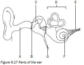
1. Identify the parts labelled B, C and F. (3)
2. Give the function of the pinna. (2)
3. Write the letter of the part which:
a) contains receptors for balance. (1)
b) equalises the pressure on either side of part B. (1)
c) transmits impulses to the brain. (1)
4. Describe how hearing occurs. (8) [16]
Answers to activity 4
1.
B -Tympanic membrane✔
C - Malleus/hammer/an ossicle✔
F - Cochlea✔(3)
2. It directs sound waves✔ into the auditory canal✔. (2)
3.
a) D✔
b) G✔
c) E✔ (3)
4.
- Sound waves are directed into the auditory canal✔ by the pinna✔.
- The sound waves make the tympanic membrane vibrate✔ and the vibrations are passed on to the ossicles✔ in the middle ear.
- The ossicles make the oval window vibrate✔ and this causes pressure waves to be set up in the inner ear.
- These vibrations also cause the organ of Corti✔ to be stimulated and it generates impulses which are sent to the cerebrum✔ along the auditory nerve✔.
- The cerebrum interprets the impulses as sound✔. (8) [16]
GENETICS GRADE 12 NOTES - LIFE SCIENCES STUDY GUIDES
GENETICS
LIFE SCIENCES
STUDY GUIDES AND NOTES
GRADE 12
- Key concepts
- Activity 1
- Genetic crosses
- Mutations
- Pedigree diagrams
- Activity 4
- Genetic engineering
- Activity 5
- Genetic counselling
- Activity 6
CHAPTER 5: GENETICS
5.1 Key concepts
Make mobile notes (see instructions on page x) to learn these key concepts.
Term | Explanation | Diagram/Additional notes |
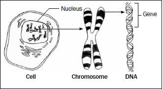
Gene | A small portion of DNA coding for a particular characteristic. |
|
Alleles | Different forms of a gene which occur at the same locus (position) on homologous chromosomes. | 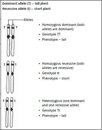 |
Genotype | Genetic composition (make-up) of an organism. | |
Phenotype | The physical appearance of an organism determined by the genotype, e.g. tall, short. | |
Dominant allele | An allele that is expressed (shown) in the phenotype when found in the heterozygous (Tt) and homozygous (TT) condition. | |
Recessive allele | An allele that is masked (not shown) in the phenotype when found in the heterozygous (Tt) condition. | |
Heterozygous | Two different alleles for a particular characteristic, e.g. Tt. | |
Homozygous | Two identical alleles for a particular characteristic, e.g. TT or tt. |
Term | Explanation | Diagram/Additional notes | |
Monohybrid cross | Only one characteristic or trait is being shown in the genetic cross. | Example: Flower colour only, e.g. yellow flower or white flower or shape of seeds only, e.g. round seeds or wrinkled seeds. | |
Complete dominance | A genetic cross where the dominant allele masks (blocks) the expression of a recessive allele in the heterozygous condition. | In this type of cross the allele for tall (T) is dominant over the allele for short (t). The offspring will therefore be tall because the dominant allele (T) masks the expression of the recessive allele (t). | 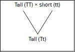 |
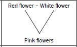
Incomplete dominance | A genetic cross between two phenotypically different parents produces offspring different from both parents but with an intermediate phenotype. | Example: If a red-flowered plant is crossed with a white-flowered plant and there is incomplete dominance - the offspring will have pink flowers (intermediate colour). |
|
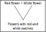
Co-dominance | A genetic cross in which both alleles are expressed equally in the phenotype. | Example: If a red-flowered plant is crossed with a white-flowered plant and there is co-dominance the offspring has flowers with red and white patches. |
|
Multiple alleles | More than two alternative forms of a gene at the same locus. | Example: Blood groups are controlled by three alleles, namely IA, IB and i. | |
Sex-linked characteristics | Characteristics or traits that are carried on the sex chromosomes. | Examples: Haemophilia and colour-blindness The alleles for haemophilia (or colour-blindness) are indicated as superscripts on the sex chromosomes, e.g. XHXH (normal female), XHXh (normal female), XhXh (female with haemophilia), XHY (normal male), XhY (male with haemophilia). | |
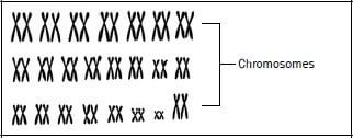
Karyotype | The number, shape and arrangement of all the chromosomes in the nucleus of a somatic cell. |
| |
Cloning | Process by which genetically identical organisms are formed using biotechnology. | Example: Dolly the sheep was cloned using a diploid cell from one parent; therefore it had the identical genetic material of that parent. | |
Genetic modification | The manipulation of the genetic material of an organism to get desired changes. | Example: The insertion of human insulin gene in plasmid of bacteria so that the bacteria produce human insulin. | |
Human genome | The mapping of the exact position of all the genes in all the chromosomes of a human. | Example: Gene number 3 on chromosome number 4 is responsible for a particular characteristic. | |
Activity 1
Choose an item from COLUMN 2 that matches a description in COLUMN 1.
Write only the letter (A to I) next to the question number (1-5), for example 6. J.
| COLUMN 1 | COLUMN 2 |
| A. Gene B. Recessive C. Haemophilia D. Dominant E. Homologous F. Genotype G. Phenotype H. Alleles I. Karyotype |
[5]
Answers to activity 1
- B✔
- H✔
- C✔
- E✔
- G✔ (5 × 1) [5]
5.2 Genetic crosses
Use the following genetic problem format or template to solve all monohybrid genetic problems: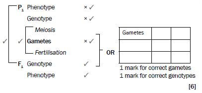
- The problem on the next page shows that a cross between a heterozygous parent (Tt) and a homozygous recessive (tt) parent produces F1 offspring that are 50% heterozygous (Tt) and 50% homozygous recessive (tt).
- A cross between a homozygous dominant (TT) parent and a homozygous recessive (tt) parent produces F1 offspring that are 100% heterozygous (Tt).
- A cross between a homozygous dominant (TT) and a heterozygous (Tt) parent produces F1 offspring that are 50% homozygous dominant (TT) and 50% heterozygous (Tt).
- A cross between two heterozygous (Tt) parents produces F1 offspring that are 25% homozygous dominant (TT), 50% heterozygous (Tt) and 25% homozygous recessive (tt).
5.2.1 Complete dominance
This refers to a genetic cross where the dominant allele masks (blocks) the expression of a recessive allele in the heterozygous condition.
The following problem represents a genetic cross which shows complete dominance:
e.g. Genetic problem 1
In humans the ability to roll the tongue is due to a dominant allele. A man who is heterozygous for tongue-rolling and a woman who cannot roll her tongue have children.
Use the symbols T and t for the alleles of the tongue-rolling characteristic and represent a genetic cross to determine the possible genotypes and phenotypes of the children. (6)
Read the problem carefully and note the following steps:
- Identify the phenotypes of the man and the woman (parents/P1), i.e. the man is a tongue-roller and the woman is a non-tongue-roller Step 1
- Identify the genotypes of the two parents, i.e. the man is heterozygous (Tt) and the woman can only be a non-tongue-roller if she is homozygous recessive for this characteristic, i.e. she must have the genotype (tt) Step 2
- The next step is to show how the alleles are separated through the process of meiosis into separate gametes, i.e. in the man the gametes (sperm) will contain either the ‘T’ allele or the ‘t’ allele.In the woman the egg can only contain the ‘t’ allele Step 3
- The next step shows that fertilisation takes place. Indicate all possible combinations of how sperm cells fuse with a possible egg cell to show the possible genotypes of the F1 generation that could arise Step 4
- Interpret the phenotypes of all the possible genotypes from the cross Step 5
Solution to genetic problem 1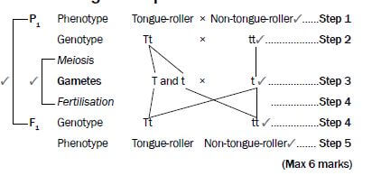
5.2.2 Incomplete dominance
This refers to a genetic cross between two phenotypically different parents producing an offspring different from both parents but with an intermediate phenotype. The following problem represents a genetic cross that shows incomplete dominance.
e.g. Genetic problem 2
A homozygous snapdragon plant with red flowers (R) was cross-pollinated with a homozygous snapdragon plant with white (W) flowers. All the plants that grew from the cross had pink flowers. Represent a genetic cross to show the possible genotypes and phenotypes of the F1 generation of plants.
Solution to genetic problem 2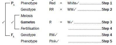
5.2.3 Co-dominance
This refers to a genetic cross in which both alleles are equally expressed in the phenotype.
The following problem represents a genetic cross which shows co- dominance.
e.g. Genetic problem 3
A plant with white flowers was cross-pollinated with a plant with red flowers. All the plants that grew from the cross had flowers with equal distribution of red and white colour. Represent a genetic cross to show the possible genotypes and phenotypes of the F1 generation of plants.
Solution to genetic problem 3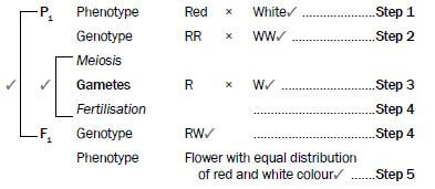
5.2.4 Inheritance of sex
The following problem represents a genetic cross which shows inheritance of sex.
e.g. Genetic problem 4
A couple has three sons and the woman is pregnant again. Show diagrammatically by means of a genetic cross what the percentage chance is of the couple having a baby
girl.
Solution to genetic problem 4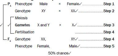
5.2.5 Inheritance of sex-linked characteristics
Sex-linked characteristics are characteristics (traits) that are carried on the sex chromosomes.
The following problem represents a genetic cross which shows the inheritance of sex-linked characteristics.
e.g. Genetic problem 5
Haemophilia is a sex-linked hereditary disease that occurs as a result of a recessive allele on the X-chromosome (Xh). A normal father and heterozygous normal mother have children. Represent a genetic cross to determine the possible genotypes and phenotypes of their children.
The alleles for haemophilia are indicated as superscripts on the sex chromosomes,
e.g. XHXH (normal female), XHXh (carrier/heterozygous normal female), XhXh (female with haemophilia), XHY (normal male), XhY (male with haemophilia).
Solution to genetic problem 5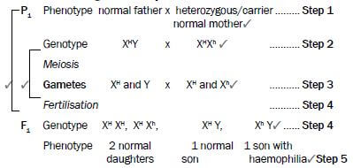
Activity 2
Question 1
Try solving this problem on your own before you look at the solution.
Fur colour in mice is controlled by a gene with two alleles. A homozygous mouse with black fur was crossed with a homozygous mouse with brown fur. All offspring had black fur. Using the symbols B and b to represent the two alleles for fur colour, show diagrammatically a genetic cross between a mouse that is heterozygous for fur colour and a mouse with brown fur. Show the possible genotypes and phenotypes of the offspring. (6)
Question 2
In rabbits the dominant allele (B) produces black fur and the recessive allele (b) produces white fur. Use a genetic cross to show the possible phenotypes and genotypes of the F1 generation for fur colour if two heterozygous rabbits are crossed. (6)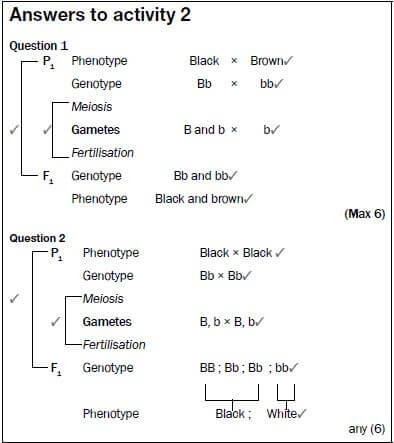
5.2.6 Dihybrid cross
- A dihybrid cross involves the inheritance of two characteristics. Mendel explained the results obtained from dihybrid crosses according to his Law of Independent Assortment.
- According to the Law of Independent Assortment, alleles of a gene for one characteristic segregate independently of the alleles of a gene for another characteristic. The alleles for the two genes will therefore come together randomly during gamete formation.
- This means that the two characteristics are transmitted to the offspring independently of one another.
- The above law only applies if the genes for the two characteristics are not on the same chromosome.
Steps you should follow in working out a dihybrid cross:
e.g. Example
In pea plants, the allele for tallness (T) is dominant and the allele for shortness (t) is recessive. The allele for purple flowers is dominant (P) and the allele for white flowers is recessive (p). Two plants, heterozygous for both tallness and purple flowers, were crossed.
STEP | What to do generally | What to do in this problem | |||||||||
Step 1 | Identify the phenotypes of the two plants for each of the two characteristics. | According to the statement of the problem, both parents are tall and have purple flowers. | |||||||||
Step 2 | Choose letters to represent the alleles for the gene responsible for each characteristic. | Use the letters, e.g. T for tall, t for short, P for purple, and p for white as provided in the question. | |||||||||
Step 3 | Write the genotypes of each parent. | According to the statement of the problem, both parents are heterozygous for each characteristic. Their genotype will therefore be TtPp. | |||||||||
Step 4 |
|
| |||||||||
Step 5 | Enter the possible gametes at the top and side of a Punnett square. | Please refer to the solution that follows. |
Step 6 |
| Please refer to the solution that follows. |
Step 7 | Determine the phenotypes of the offspring from the genotypes obtained in the punnet square. | Please refer to the solution that follows. |
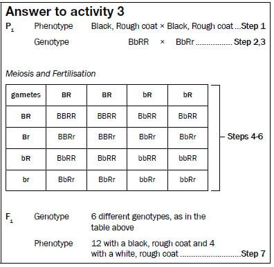
Solution to the problem
P1 Phenotype Tall, Purple × Tall, Purple................... Step 1
Genotype TtPp × TtPp......................... Step 2,3
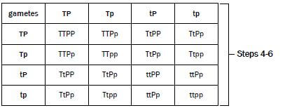
F1 Genotype 9 different genotypes, as in the table above
Phenotype 9 tall, purple flowered plants (T-P-);
3 short, purple flowered plants (ttP-);
3 tall, white flowered plants (T-pp), and
1 short, white flowered plant (ttpp) . Step 7
Activity 3
Question
In hamsters, the allele for black coat colour (B) is dominant over the allele for white coat colour (b). The allele for rough coat (R) is dominant over the allele for smooth coat (r). If you cross a hamster that is heterozygous black and homozygous rough, with one that is heterozygous black and heterozygous rough, what will be the phenotypes and genotypes of the offspring? (Use the steps 1-7 to arrive at an answer).

5.3 Mutations
A mutation is any sudden unexpected change in the genetic structure of a cell. Mutations occur suddenly and randomly and may be caused by many environmental agents such as X-rays and certain chemicals.
Mutations may be harmful or harmless to the organism in which they occur. Harmful mutations cause changes in DNA that can cause errors in protein sequencing, that can result in partially or completely non-functional proteins. Harmless mutations have no effect on the structure or functioning of the organism. Useful mutations can be advantageous to the organism and they are passed on from parent to offspring.
Gene mutations are mutations that affect a single or a few base pairs in just a single gene, while Chromosomal aberrations refer to changes in the normal structure or number of chromosomes.
Mutations result in new genotypes as we move from one generation to the next. This leads to variation within a species.
Gene mutations can cause genetic disorders:
- Haemophilia: Absence of the protein needed for the formation of blood clots due to a mutant gene.
- Colour blindness: Absence of the proteins that make up either the red or green cones/photoreceptors in the eye.
- Albinism: Absence of the protein that forms the pigment melanin.
Chromosomal aberrations e.g. Down syndrome is where there is an extra chromosome (47 instead of 46) in the zygote.
5.4 Pedigree diagrams
A pedigree diagram is used to study the inheritance of characteristics in a family over a number of generations. A pedigree diagram is also called a family tree.
Remember the following steps when interpreting pedigree diagrams:
Step 1 Study any key and opening statement/s and look for dominant and recessive characteristics and phenotypes.
Step 2 Write in the phenotypes of all the individuals as given in the problem.
Step 3 Fill in the genotype of all the individuals with the recessive condition - it must have two recessive alleles (two lower case letters, e.g. ff).
Step 4 For every individual in the diagram that has the recessive condition, it means that each allele was obtained from each of the parents. Work backwards and fill in one recessive allele for each parent.
Step 5 If the parents showed the dominant characteristic, fill in the second letter which represents the dominant allele (a capital letter, e.g. F).
Step 6 Any other individual showing the dominant characteristic will most likely be homozygous dominant (FF) or heterozygous dominant (Ff).
Activity 4
The pedigree diagram in Figure 5.1 shows inheritance of eye colour in humans over three generations of a family. Brown eye colour (B) is dominant over blue eye colour (b). Study the diagram and then answer the questions that follow.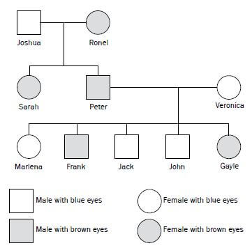
Figure 5.1 Pedigree diagram showing inheritance of eye colour
Note the following in the pedigree diagram on page 38:
- Squares represent males and circles represent females.
- The horizontal line between a square (Joshua) and a circle (Ronel) shows that they have mated.
- The vertical line flowing from the horizontal line represents the offspring (Sarah and Peter) of the two parents (Joshua and Ronel).
- Brown eye colour (B) is dominant over blue eye colour (b) - stated in problem Step 1
- Joshua, Jack and John are males with blue eyes.
- Veronica and Marlena are females with blue eyes.
- Peter and Frank are males with brown eyes. Step 2
- Ronel, Sarah and Gayle are females with brown eyes.
- Joshua, Veronica, Marlena, Jack and John will have the genotype ‘bb’. The recessive characteristic only shows up in the homozygous condition Step 3
- Example: The genotype of Peter is ‘Bb’ - working backwards from the offspring Marlena or Jack or John who are homozygous recessive.
This means that one of the recessive alleles of Marlena, Jack and John, i.e. ‘b’, must have come from parent Peter and the other one from parent Veronica Steps 4 and 5 - Ronel could be homozygous dominant (BB) or heterozygous dominant (Bb) Step 6
Questions
1. How many members of the family have blue eyes? (1)
2. Is Veronica homozygous or heterozygous for eye colour? (1)
3. Write down the genotype of:
a) Joshua (2)
b) Ronel (2)
c) Frank (2)
4. If Frank marries a woman with the same genetic composition as Sarah, what is the percentage probability of them having a child with brown eyes? (1) [9]
Answers to activity 4
1. 5✔ (1)
2. Homozygous✔(1)
3. a) bb✔✔ (2)
b) BB/Bb✔✔ (2)
c) Bb✔✔ (2)
4. 75%✔
5.5 Genetic engineering
Genetic engineering is the process whereby the genes on the DNA are changed, transferred or manipulated to produce a different organism.
Activity 5
Question
State FOUR disadvantages and FOUR advantages of genetic engineering. [8]
Answer to activity 5
Four disadvantages of genetic engineering:
- Expensive✔/research money could be used for other needs
- Interfering with nature✔/immoral
- Potential health impacts✔
- Unsure of long-term effects✔ (4)
Four advantages of genetic engineering:
- Production of medication/resources cheaply✔
- Control pests with specific genes inserted into a crop✔
- Using specific genes to increase crop yields✔/food security
- Selecting genes to increase shelf-life of plant products✔ (4) [8]
5.6 Genetic counselling
Couples with a risk of a genetic disease can undergo genetic counselling to enable them to make informed decisions on whether they want to have children or not.
Activity 6
Question
A young couple wants to have a child, but they are aware of a serious genetic disorder in one of their families that could be carried through to their offspring. State THREE benefits of genetic counselling in this case. [3]
Answer to activity 6
Three benefits of genetic counselling:
- To be given advice on the risk of transferring the defective gene✔/ to find the probability of passing on the defective gene to the offspring
- To be given an explanation of the procedure involved in DNA testing✔
- To be given an explanation of the results of DNA testing✔ [3]
REPRODUCTION GRADE 12 NOTES - LIFE SCIENCES STUDY GUIDES
REPRODUCTION
LIFE SCIENCES
STUDY GUIDES AND NOTES
GRADE 12
- Male reproductive system
- Activity 1
- Female reproductive system
- Activity 2
- Puberty
- Menstrual cycle
- Hormonal control of the menstrual cycle
- Activity 3
- Development of the foetus
- Activity 4
CHAPTER 4:REPRODUCTION
4.1 Male reproductive system
Figure 4.1 below shows the different parts of the male reproductive system and their functions.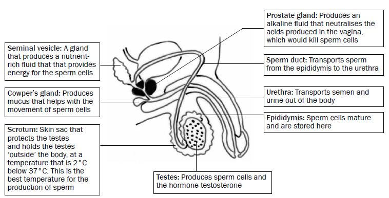
Figure 4.1 Structure of the male reproductive system
Functions of testosterone
The testes produce the hormone testosterone, which has the following functions:
- Development of male secondary sexual characteristics, such as beard, pubic hair, deep voice and a muscular body.
- Stimulates the maturation of sperm cells.
Structure of a sperm cell
Figure 4.2 below shows the different parts of a sperm cell and their functions.
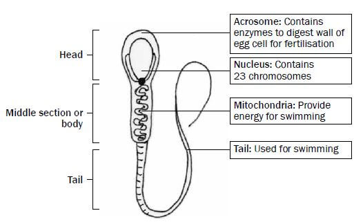
Figure 4.2 Structure of a sperm cell
Activity 1
Questions
- Name the accessory glands of the male reproductive system and give ONE function of each. (10)
- Name the organ where testosterone is produced. (1)
- Give TWO functions of testosterone. (2)
- Name all the parts of the sperm cell that are responsible for movement. State what the function of each part is. (4)
- Explain the role of the nucleus of the sperm cell in fertilisation. (3) [20]
Answers to activity 1
- Seminal vesicle ✔ produces a fluid that contains nutrients ✔ for the sperm cells, so that they have energy to swim.✔
Prostate gland ✔ produces an alkaline fluid ✔ that neutralises acids ✔ produced in the vagina, so that sperm cells are protected.✔
Cowper’s gland✔ produces mucus✔ that helps with the movement✔of sperm cells. (10) - Testes✔ (1)
- Testosterone is responsible for the development of male secondary sexual characteristics✔ and it stimulates the maturation of sperm cells.✔ (2)
- Mitochondria✔ provide energy for swimming.✔ Tail✔ moves in a whip-like fashion to propel the sperm cell forwards.✔ (4)
- The nucleus contains 23 chromosomes (n)✔, and fuses with the nucleus of an egg cell, which also contains 23 chromosomes (n)✔. The result is a zygote with
46 chromosomes (2n).✔ (3) [20]
4.2 Female reproductive system
Figure 4.3 below shows the different parts of the female reproductive system and their functions.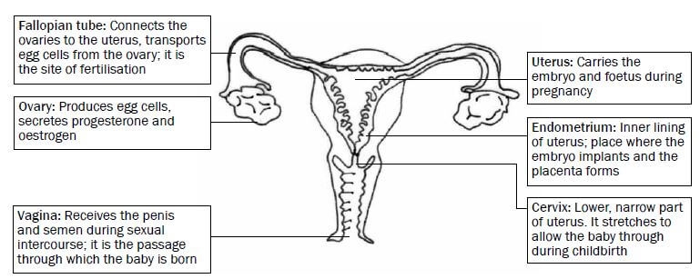
Figure 4.3 Structure of the female reproductive system
Activity 2
Questions
Provide the correct biological term for the following definitions.
- The inner lining of the uterus (1)
- Tube that connects the ovaries to the uterus (1)
- The structure that produces female hormones (1)
- The part where development of the embryo/foetus normally takes place in humans (1) [4]
Answers to activity 2
- Endometrium✔
- Fallopian tube✔
- Ovary/placenta✔
- Uterus✔ [4]
4.3 Puberty
Puberty is the period in humans in which they experience physical changes in their bodies in order to be capable of sexual reproduction.
Puberty in males | Puberty in females |
Stimulated by testosterone | Stimulated by oestrogen |
Growth of male sex organs | Growth of female sex organs |
Start of the production of sperm cells | Start of the menstrual cycle and production of ova |
Growth of pubic hair, facial hair and body hair | Growth of pubic hair |
Development of muscles and deepening of voice | Growth and development of breasts and widening of hips |
4.4 Menstrual cycle
The series of diagrams in Figure 4.4 below shows the events occurring in the ovary (ovarian cycle) and uterus (uterine cycle) during the menstrual cycle. The days are not exact, but are averages.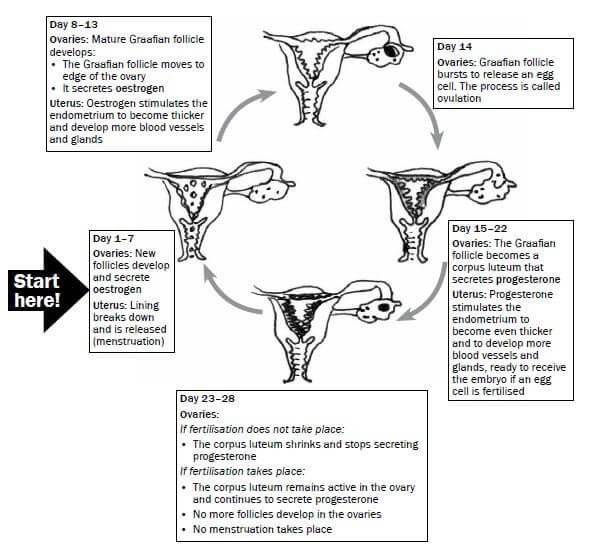
Figure 4.4 The menstrual cycle
4.5 Hormonal control of the menstrual cycle
The graph in Figure 4.5 below shows changes in the ovary, uterus and in the level of hormones during a 28-day menstrual cycle.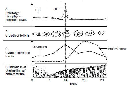
Figure 4.5 Hormonal regulation of the female reproductive cycle
The hormonal changes that take place at A, B, C and D in the graph in Figure 4.5 above are explained in Table 4.1 below.
| A | B | C | D | |
| Day 0-11 | Pituitary gland produces FSH which stimulates development of the follicle. | Follicle is developing to become a Graafian follicle containing an egg cell. | Oestrogen levels increase as the hormone is produced by the follicle. | Thickness of endometrium increases from day 7 (after menstruation has ended) as a result of oestrogen. |
| Day 11- 17 | FSH and LH (produced by the pituitary gland) levels are highest around day 14. | Follicle development is completed as a result of the influence of FSH by day 14. Ovulation is stimulated by high levels of FSH and LH on day 14. LH then stimulates the development of the corpus luteum. | Oestrogen levels reach a maximum towards day 14 until ovulation takes place, but then start to decrease because the Graafian follicle stops functioning. | Endometrium thickens further. |
| Day 17-28 | LH levels decrease and then remain constant to maintain the corpus luteum. | Corpus luteum produces progesterone. Corpus luteum gradually disintegrates since fertilisation does not take place. | Oestrogen levels increase again and then decrease towards the end of the cycle. Progesterone levels increase towards day 21. Progesterone levels decrease when corpus luteum disintegrates and stops functioning. | Progesterone prepares endometrium fully for pregnancy. Decreased progesterone levels from around day 21 cause endometrium to shed after day 28 by menstruation since no fertilisation took place. |
Table 4.1 Hormonal changes during the menstrual cycle
Activity 3
Study Figure 4.6 below and answer the questions that follow.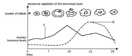
Figure 4.6 Hormonal changes during the menstrual cycle
- Name the hormones A and B. (2)
- Give reasons for your answers in question 1. (2)
- What event occurs on day 14? (1)
- Name the other two hormones involved in this cycle. (2)
- Did fertilisation occur during the cycle shown in Figure 4.6? (1)
- Explain your answer in question 5. (2) [10]
Answers to activity 3
- A - Oestrogen✔ B - Progesterone✔ (2)
- A: The Graafian follicle secretes oestrogen✔/Oestrogen reaches its maximum level before ovulation.✔
B: The corpus luteum produces progesterone✔/Progesterone reaches its maximum level after ovulation.✔ (2) - Ovulation✔ (1)
- LH✔ and FSH✔ (2)
- No✔ (1)
- Progesterone levels decrease✔ towards the end of the cycle.
The corpus luteum decreases✔ in size. (2) [10]
HINT:
Here is a hint to help you to remember the names of the two hormones:
- O stands for Oestrogen and when it is high, Ovulation occurs.
- P stands for Progesterone and when it remains high, there is a Pregnancy.
4.6 Development of the foetus
Figure 4.7 below shows the stages in the development of the foetus.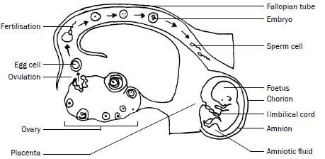
Figure 4.7 Stages in the development of the foetus
Explanation of Figure 4.7
- In the ovary a mature Graafian follicle bursts (usually on day 14 of the menstrual cycle) and releases an egg cell. This process is called ovulation.
- Fertilisation takes place high up in the fallopian tube. The egg cell (containing 23 chromosomes) and sperm cell (containing 23 chromosomes) fuse to form a zygote (containing 46 chromosomes).
- The zygote divides by mitosis to form a morula, then a blastocyst, and finally an embryo as it moves down the Fallopian tube.
- It takes about 5 to 7 days for the embryo to reach the uterus.
- In the uterus the embryo settles on the endometrium and sinks into it, embedding itself in the endometrium. This process is called implantation.
- After implantation, the embryo produces many finger-like structures called villi from the outer membrane of the embryo, which is known as the chorion.
- The villi grow into the tissue of the uterus to form a placenta.
- The placenta is attached to the embryo by the umbilical cord. It has 2 umbilical arteries (which carry deoxygenated blood from the embryo towards the placenta) and 1 umbilical vein (which carries oxygenated blood from the placenta to the embryo).
- The embryo is enclosed in a fluid-filled sac called the amnion. The fluid is called the amniotic fluid.
- After about 8 weeks, the embryo develops structures such as limbs and all the organs of the body. Now it is called a foetus.
- Gestation is the period between fertilisation and the birth of the baby. It usually lasts for a period of 9 months (39-40 weeks).
- The stages involved in the natural birth process are:
- Dilation of the cervix (labour)
- Expulsion of the foetus.
- Delivery of the afterbirth (placenta) and extra-embryonic membranes.
Activity 4
Questions
- On which day of the menstrual cycle does ovulation usually take place? (1)
- What happens to the Graafian follicle after ovulation? (1)
- Name the TWO hormones that are released by structures in the ovaries. (2)
- Give THREE functions of the amniotic fluid. (3)
- Give TWO substances that can move from the mother to the foetus through the placenta. (2)
- Give TWO substances that can move from the foetus to the mother through the placenta.
Answers to activity 4
- Day 14✔ (1)
- It changes into a corpus luteum.✔ (1)
- Oestrogen✔ and progesterone.✔ (2)
- The amniotic fluid protects the foetus against shock✔, drying out✔ and temperature changes.✔ (3)
- Oxygen✔, nutrients✔ (amino acids, glucose, other sugars), viruses✔ and drugs✔ (2)
- Carbon dioxide✔ and waste products✔ (urea). (2) [11]
REPRODUCTION IN VERTEBRATES GRADE 12 NOTES - LIFE SCIENCES STUDY GUIDES
REPRODUCTION IN VERTEBRATES
LIFE SCIENCES
STUDY GUIDES AND NOTES
GRADE 12
CHAPTER 3: REPRODUCTION IN VERTEBRATES
Different groups in the animal kingdom have different strategies to maximise reproductive success in different environments. These are a few of the strategies used by vertebrates.
Strategy | How it works? | What is its advantage? |
External Fertilisation | The sperm fertilises the egg outside the body of the female, usually in water. | Water prevents the eggs from drying out and allows the sperm to swim towards the egg. |
Internal Fertilisation | The male deposits its sperm inside the reproductive organs of the female and fertilisation occurs inside the female’s reproductive organs. | Allows terrestrial animals to reproduce in a dry environment without the need for water. |
Ovipary | Eggs are laid and hatching takes place outside the mother’s body. | Egg provides nutrition for the developing embryo and protects the embryo. A shelled egg frees these animals from the need to reproduce in water. |
Vivipary | The young develop inside the uterus of the mother after the eggs are fertilised internally. | More efficient development of the embryo as nutrients are received for a longer period from the mother’s blood through a placenta. Embryo is protected in the body of the mother. |
Ovovivipary | Young develop from eggs that are fertilised internally and retained within the mother's body after fertilisation until they hatch. | Embryos obtain their nutrients from the egg yolk. The eggs are protected from predators until hatching occurs. |
Amniote Egg | Embryo protected by the shell of the egg; egg consists of many extraembryonic membranes that serve different functions. | Amniote egg protects embryo from dehydration. Yolk sac provides nutrition, Allantois for excretion, chorion for gas exchange. |
Precocial Development | Hatchlings are quite well-developed when they hatch - eyes open, able to move, able to feed. Brain size and intelligence remains the same throughout their lives. | Hatchlings are more prepared to handle the challenges of the environment; More independent. |
Altricial Development | Hatchlings are poorly-developed when they hatch. Unable to feed on their own, cannot move. Brain size and intelligence increases a lot after hatching. | Parental care afforded to protect the young from predators. |
Parental Care | Parental care offered through building of nests, protecting the eggs, protecting the young, teaching the young. | Increases chances of survival of the young. |
Activity 1
Questions
Indicate whether each of the statements in COLUMN I applies to A ONLY, B ONLY, BOTH A and B or NONE of the items in COLUMN II. Write A only, B only, both A and B, or none next to the question number (1 to 5).
COLUMN I | COLUMN II |
1. Oviparous | A Eggs are produced B Eggs are always incubated by the female |
2. Ovoviviparous | A Eggs incubated in nests B Eggs incubated in the female’s body |
3. Precocial | A Small, helpless offspring born B Intense parental care required |
4. Viviparous | A Gestation period required B Live offspring born |
5. Altricial | A Intense parental care required B Offspring can look after themselves |
(5 × 2 ) [10]
Answers to activity 1
- A only (B is wrong, because some animals, like insects, simply lay their eggs and do not incubate them. In some birds both the male and female incubate the eggs)
- B only (A is wrong, because the eggs are not released from the female’s body)
- None (Precocial animals are born quite well-developed, they can live independently from their parents and find their own food, so parental care is not required)
- Both A and B
- A only (B is wrong, because altricial animals are born small and helpless. They cannot look after themselves or find their own food. Their parents must look after them, protect them and feed them.) (5 × 2) [10)
MEIOSIS GRADE 12 NOTES - LIFE SCIENCES STUDY GUIDES
MEIOSIS
LIFE SCIENCES
STUDY GUIDES AND NOTES
GRADE 12
What the chapter entails:
- What is meiosis?
- The process of meiosis in animal cells
- The significance of meiosis
- Abnormal meiosis
- Differences between meiosis I and meiosis II
- Worked example
- Activity 1
CHAPTER 2: MEIOSIS
2.1 What is meiosis?
Meiosis is a type of cell division whereby a diploid cell (somatic cell) undergoes two cell divisions, and divides to form four dissimilar haploid cells (sex cells). Diploid cells have two sets of chromosomes, where each chromosome has a homologous partner. Haploid cells only have one set of chromosomes. Chromosomes in haploid cells have no homologous
partners.
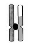
Before meiosis begins (during interphase), DNA replication takes place. The result is two sets of chromosomes consisting of two identical chromatids joined together with a centromere. This is shown in Figure 2.1 (below).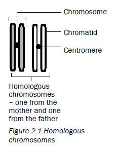
2.2 The process of meiosis in animal cells
Meiosis is the type of cell division used to produce gametes or sex cells (sperm and egg cells). A cell undergoing meiosis will divide twice - the first division is meiosis I and the second is meiosis II. | 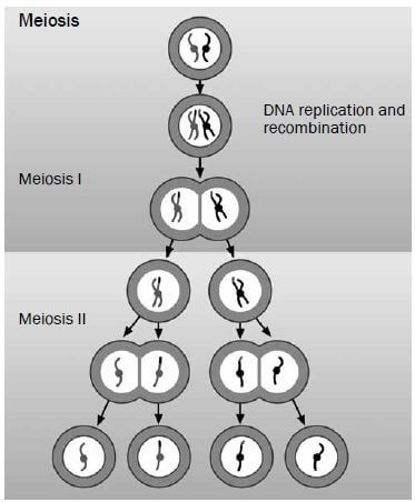 |
2.2.1 First meiotic division
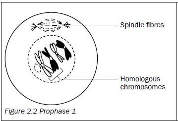
| Prophase 1
|
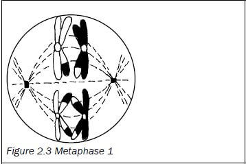
| Metaphase 1
|
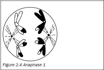
| Anaphase 1
|
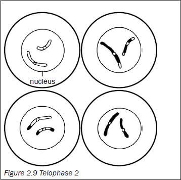
| Telophase 1
|

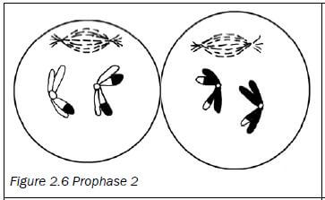
2.2.2 Second meiotic division
| Prophase 2
|
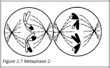
| Metaphase 2
|
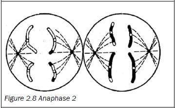
| Anaphase 2
|
| Telophase 2
|
An easy way to remember the events of meiosis is to use the word mnemonic IPMAT.
Letter | Phase | Event |
I | Interphase | I for in between: The part of the life cycle of the cell that is in between cell divisions. |
P | Prophase | P for preparation: The chromosomes prepare for meiosis by untangling and becoming clearly visible. Crossing over also takes place. |
M | Metaphase | M for middle: The chromosomes move to the ‘middle’ (equator). |
A | Anaphase | A for apart: The chromosomes/chromatids move apart/move to the poles. |
T | Telophase | T for terminal: The final phase of meiosis I/ meiosis II. |
2.3 The significance of meiosis
There are two reasons why meiosis is important.
- It reduces the number of chromosomes by half, in other words from diploid to haploid. This ensures that sex cells have half the number of chromosomes of other somatic cells so that when fertilisation occurs the zygote formed has the correct number of chromosomes. It balances the doubling effect of fertilisation.
- Crossing over introduces genetic variation. Genetic variation results in offspring that are better adapted to a particular environment and ensures that they will have a better chance of survival.
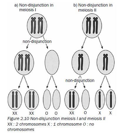
2.4 Abnormal meiosis
- Sometimes mistakes occur during the process of meiosis.
- This can happen in Anaphase 1 where the homologous chromosomes may not separate. Also called non-disjunction. (see above)
- It can also happen in Anaphase 2 when there is non-disjunction of the sister chromatids.
- If there is non-disjunction of chromosome pair 21 in humans it leads to the formation of an abnormal gamete with an extra copy of chromosome 21.
- If there is fusion between a normal gamete and an abnormal gamete (with extra copy of chromosome 21) it leads to Down Syndrome.
2.5 Differences between meiosis I and meiosis II
Meiosis I | Meiosis II |
The chromosomes arrange at the equator of the cell in homologous pairs. | Chromosomes line up at the equator of the cell individually. |
Whole chromosomes move to opposite poles of the cell. | Daughter chromosomes/chromatids move to opposite poles of the cell. |
Two cells form at the end of this division. | Four cells are formed at the end of this division. |
The chromosome number is halved during meiosis I. | The chromosome number remains the same during meiosis II. |
Crossing over takes place. | Crossing over does not take place. |
Table 2.1 The differences between meiosis I and meiosis II
Worked example
Study the diagrams below of two stages of meiosis then answer the questions that follow.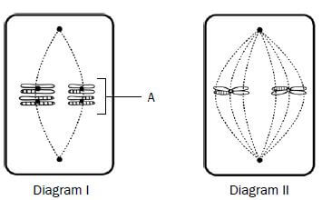
Figure 2.11 Two stages of meiosis
- State ONE visible reason in Diagram I which indicates that meiosis is taking place. (1)
- How many chromosomes would be present in each daughter cell at the end of meiosis in this cell? (1)
- Describe what takes place in the cell after the phase shown in Diagram I. (3)
- Tabulate TWO visible differences between the phases of meiosis shown in Diagrams I and II. (5) [10]
Answers to worked example
- The chromosomes are lined up at the equator of the cell in their homologous pairs.✔
OR
The chromosomes show evidence of crossing over.✔ (1) - Two ✔ chromosomes. (1)
- The next phase is Anaphase 1. The spindle fibres contract.✔ (shorten) and pull each chromosome ✔ of each chromosome pair to opposite poles ✔ of the cell.
(5) [10]Diagram I (metaphase 1)
Diagram II (metaphase 2)
1. Chromosomes are lined up at the equator in homologous pairs.✔
1. Chromosomes are lined up at the equator individually.✔
2. Four chromosomes are present✔
2. Two chromosomes are present✔
Activity 1
Question 1
Give the correct word or term for each of the statements or definitions provided below.
1.1 | The structure that joins the two halves of a double-stranded chromosome (1) |
1.2 | A pair of chromosomes, one inherited from each parent, that have the same genes at the same locus (1) |
1.3 | A single-stranded chromosome formed during Anaphase 2 (1) |
1.4 | The point of contact between two chromosomes of a homologous pair during crossing over (1) |
1.5 | One half of a double-stranded chromosome (1) |
1.6 | The phase in meiosis where crossing over occurs (1) [6] |
Answers to question 1
1.1 Centromere✔(1)
1.2 Homologous chromosomes✔ (1)
1.3 Daughter chromosome/chromatid✔ (1)
1.4 Chiasma✔/chiasmata✔ (1)
1.5 Chromatid✔ (1)
1.6 Prophase 1✔ (1) [6]
Question 2
Figure 2.12 (right) represents a process taking place during meiosis. Study the diagram and answer the questions that follow.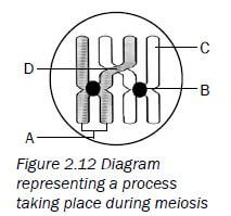
2.1 Provide labels for parts A, B, C and D. (4)
2.2 Name the process in meiosis that is illustrated in Figure 2.12. (1)
2.3 State ONE importance of the process you named in question 2.2. (2)
2.4 Draw a diagram of the structure labelled A to show its appearance immediately after the process you named in question 2.2. (2) [9]
Answers to question 2
2.1
A - Chromosome✔
B - Centromere✔
C - Chromatid✔
D - Chiasma✔/chiasmata (4)
2.2 Crossing over✔ (1)
2.3 It introduces genetic✔ variation✔ (2)
2.4
- A double-stranded chromosome with the strands joined by a centromere✔
- There is evidence of crossing over.✔ (2)

Question 3
Figure 2.13 (below) represents an animal cell in a phase of meiosis. Study the diagram and answer the questions that follow.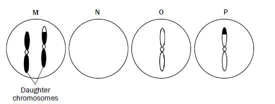
3.1 State whether the phase of meiosis shown in Figure 2.13 is meiosis I or meiosis II. (1)
3.2 Give ONE visible reason for your answer in question 3.1. (1)
3.3 Identify the parts labelled A and B. (2)
3.4 How many chromosomes:
- were present in the parent cell before meiosis began? (1)
- will be present in each cell at the end of meiosis? (1)
3.5 State ONE place in a human female where meiosis would take place. (1)
3.6 Could the cell represented in Figure 2.13 be that of a human? (1)
3.7 Explain your answer to question 3.6. (2)
3.8 Give TWO reasons why meiosis is biologically important. (2)
3.9 Give the term for the situation when some of the chromosomes do not separate correctly during the phase shown in Figure 2.13. (1) [13]
Answers to question 3
3.1 Meiosis II✔ (1)
3.2 Daughter chromosomes/chromatids are being pulled to the opposite poles✔ (1)
3.3
A - Spindle fibre✔
B - Cell membrane✔ (2)
3.4
a) 8✔
b) 4✔ (2)
3.5 Ovaries✔ (1)
3.6 No✔ (1)
3.7 There are only 4 chromosomes present3 instead of 23.✔ (2)
3.8
- It introduces genetic variation.✔
- It balances the doubling effect of fertilisation as it halves the number of chromosomes in the sex cells.✔ (2)
3.9 Non-disjunction✔ (1) [13]
Question 4
The diagram below shows the nuclei of the four cells that resulted from meiosis of chromosome pair 21 in a woman.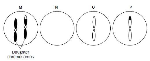
Figure 2.14: Diagram that shows the nuclei of four cells resulted from meiosis
4.1 Explain why nucleus N does NOT have a chromosome pair 21. (2)
4.2 Name and explain the disorder that will result if diagram M represents an egg cell that fuses with a normal sperm cell. (3) [5]
Answers to question 4
4.1 During Anaphase 1 the chromosome pair 21 does not separate✔/ non-disjunction. Gamete M will have an extra copy of chromosome number 21 and therefore gamete N does not have a copy of chromosome 21✔ (2)
4.2 Down syndrome✔/Trisomy 21 if gamete M fuses with normal sperm having 1 copy of chromosome 21 ✔ the resulting zygote will have 3 copies of chromosome 21✔ (3) [5]
For more papers on Meiosis visit https://www.elimuza.com/
NUCLEIC ACIDS GRADE 12 NOTES - LIFE SCIENCES STUDY GUIDES
NUCLEIC ACIDS
LIFE SCIENCES
STUDY GUIDES AND NOTES
GRADE 12
Content Covered
- The structure of DNA and RNA
- Differences between DNA and RNA
- DNA replication
- DNA profiling
- Activity 1
- Protein synthesis
- Activity 2
CHAPTER 1:Nucleic acids
1.1 The structure of DNA and RNA
- Two kinds of nucleic acids are found in a cell, namely DNA and RNA.
- These two nucleic acids are made of building blocks (or monomers) called nucleotides.
- Figure 1.1 (right) shows what a nucleotide looks like.
Table 1.1 (below) shows the nitrogenous bases of DNA and RNA.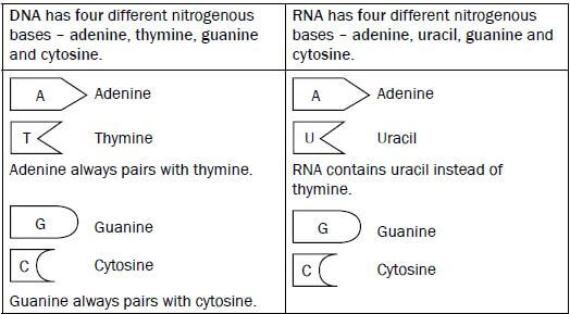
Table 1.1 Nitrogenous bases of DNA and RNA
- P - Phosphate group
- S - Deoxyribose or ribose sugar
- N - Nitrogenous base (adenine, thymine, guanine, cytosine or uracil)
Figure 1.1 below: A nucleotide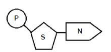
Figure 1.2 below shows the structure of DNA and RNA. Study the diagrams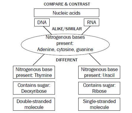
in Figure 1.2, and then read the information in the boxes below the diagrams to find out how to tell a DNA molecule from an RNA molecule.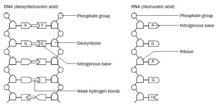 Figure 1.2 The structure of DNA and RNA
How to recognise a DNA molecule | How to recognise an RNA molecule |
|
|
|
|
| |
|
1.2 Differences between DNA and RNA
Table 1.2 below summarises the differences between DNA and RNA molecules.
DNA | RNA |
1. Double-stranded molecule | 1. Single-stranded molecule |
2. Contains deoxyribose (sugar) | 2. Contains ribose (sugar) |
3. Contains the nitrogenous base, thymine | 3. Contains the nitrogenous base, uracil |
Table 1.2 The differences between DNA and RNA
1.3 DNA replication
DNA replication takes place at interphase before mitosis or meiosis begins. DNA replication is the process during which a DNA molecule makes an exact copy (replica) of itself. This is shown in Figure 1.3 below.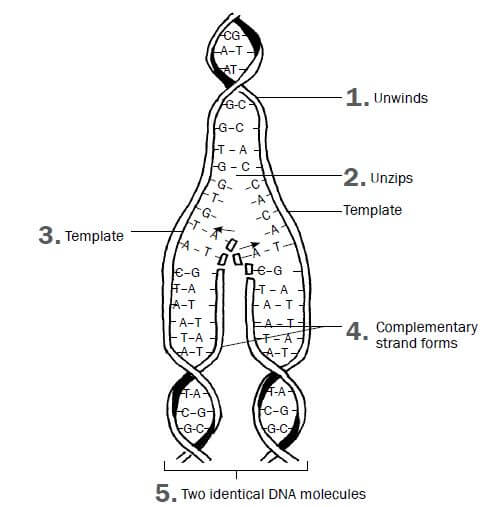
Figure 1.3 DNA replication
- The double helix CG unwinds.
- Weak hydrogen bonds between nitrogenous bases break and two DNA strands unzip (separate).
- Each original DNA strand serves as a template on which its complement is built.
- Free nucleotides build a DNA strand onto each of the original two DNA strands by attaching to their complementary nitrogenous bases (A to T and C to G).
- This results in two identical DNA molecules. Each molecule consists of one original strand and one new strand.
Significance of DNA replication
DNA replication is important because it:
- Doubles the genetic material so it can be shared between the resulting daughter cells during cell division.
- Results in the formation of identical daughter cells during mitosis.
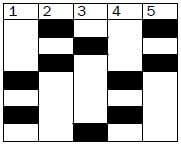
1.4 DNA profiling
Every person except identical twins has her/his own unique DNA profile. It can be described as an arrangement of black bars representing DNA fragments of the person.
It is used to:
- Identify criminals
- Identify dead bodies
- Identify relatives
- Identify paternity
Activity 1
- A DNA molecule contains 600 nitrogen bases. If 20% of this is adenine, determine the number of each nitrogen base in the DNA molecule. (3)
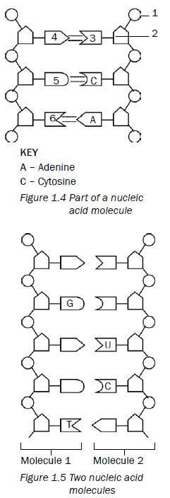
- Figure 1.4 (below) represents part of a nucleic acid molecule. Study the diagram and answer the questions that follow.
2.1 Identify the nucleic acid shown in Figure 1.4. (1)
2.2 Label the following: (drawing below)- Part 1 (1)
- Part 2 (1)
- The nitrogenous bases 4, 5 and 6 (3)
2.3 What is the collective name for the parts numbered 1, 2 and 3?(1)
- Questions 3.1 and 3.2 are based on Figure 1.5 (below). This is a diagrammatic representation of a part of two different nucleic acid molecules found in the cells of organisms during a stage in the process of protein synthesis.
3.1 Name the molecules 1 and 2. (2)
3.2 Give a reason for your answer in question 3.1. (2) - The result of profiling various DNA samples in a criminal investigation is shown below
 (Key below}
4.1 Was suspect X or suspect Y involved in the crime? (1)
4.2 Does the DNA of the suspect (from answer 4.1) match the first or second sample? (2) [17]
 (Key below}
| Key for question 4: | Drawings for Q2 and Q3 above |
|  |
Answers to activity 1
- 20% adenine = 20% thymine✔ 30% cytosine✔= 30% guanine✔
20 × 600 = 120A = 120T 30 × 600 = 180C = 180G (3)
100 100 - 2.1 DNA✔
2.2- Phosphate✔ group (1)
- Deoxyribose✔sugar (1)
- 4 - adenine (A)✔
6 - thymine✔
5 - guanine (G)✔ (3)
2.3 Nucleotide✔ (1)
- 3.1 1 - DNA 2 - mRNA/RNA✔ (2)
3.2 DNA contains the nitrogenous base thymine (T).✔
RNA contains the nitrogenous base uracil (U).✔ (2) - 4.1 Suspect X was involved. ✔(1)
4.2 The DNA of suspect X matches with the second sample. ✔✔ (2) [17]
1.5 Protein synthesis
Protein synthesis is the process by which proteins are made in each cell of an organism to form enzymes, hormones and new structures for cells.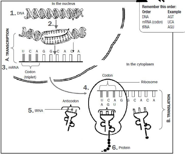
Figure 1.6 The process of protein synthesis
There are two main processes involved in protein synthesis, namely transcription and translation. They are labelled as A and B in Figure 1.6 above.
- Note that the numbers on the diagram correspond with the description below.
A Transcription (takes place in the nucleus)
- DNA unwinds and splits.
- One DNA strand acts as a template for forming mRNA.
- Free nucleotides arrange to form mRNA according to the DNA template. This process is called transcription.
- The mRNA leaves the nucleus through the nuclear pores. Stage
B now takes place when mRNA in the cytoplasm attaches to the ribosome.
B Translation (takes place in the cytoplasm on the ribosome)
5. Each tRNA brings a specific amino acid to the mRNA. This is called translation.
6. The amino acids are linked together to form a particular protein.
The diagram shown in Figure 1.6 (on page 5) may appear in exam questions in different ways. Do not let the different representations confuse you. Just try to identify the
following components by looking for the features listed here:
- DNA - double-stranded; look for presence of thymine; found in nucleus only.
- Nuclear membrane - has nuclear pores through which mRNA moves.
- mRNA - single-stranded; look for presence of uracil; contains a triplet of bases (codon) found in nucleus and cytoplasm.
- Ribosome - usually mRNA attached to it.
- tRNA - contains a triplet of bases (anticodon); look for attached amino acid.
Activity 2
Question 1
Study Figure 1.7 (below), which shows the process of protein synthesis, and answer the questions.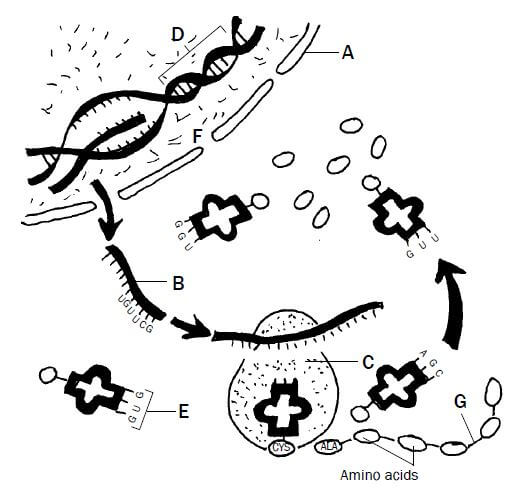
Figure 1.7 Protein synthesis
1.1 Label structures A, B and D. (3)
1.2 State ONE function of molecule D. (1)
1.3 Which stage of protein synthesis takes place at F? (1)
1.4 Identify organelle C. (1)
1.5 Name and describe the stage of protein synthesis that takes place at organelle C. (7)
1.6 Write down the codon of anticodon E from top to bottom. (1)
1.7 Name the type of bond (labelled G) between the amino acids. (1) [15]
Answers to question 1
1.1
A - Nuclear membrane✔
B - mRNA✔
D - DNA✔ (3)
1.2 Carrying hereditary characteristics from parents to their offspring✔ OR Controls the synthesis (manufacturing) of proteins✔ (1)
1.3 Transcription✔ (1)
1.4 Ribosome✔ (1)
1.5 Translation✔
- The mRNA strand from the nucleus becomes attached✔to a ribosome with its codons exposed
- each tRNA molecule carrying a specific amino acid✔
- according to its anticodon✔
- matches up with/complements the codon of the mRNA✔
- so that the amino acids are placed in the correct sequence✔
- adjacent amino acids are linked✔
- to form a protein✔ (7)
1.6 CAC✔ (the anticodon is GUG, so the complementary codon is CAC) (1)
1.7 Peptide Bond (1) [15]
Question 2
Table 1.3 below shows the DNA base triplets that code for different amino acids.
Amino acid | Base triplet in DNA template |
Leu (leucine) | GAA |
His (histidine) | GTA |
Lys (lysine) | TTT |
Pro (proline) | GGG |
Ala (alanine) | CGA |
Trp (tryptophan) | ACC |
Phe (phenylalanine) | AAA |
Gly (glycine) | CCT |
Table 1.3 Different amino acids and their DNA base triplets
The following is a part of a sequence of amino acids that forms a particular protein molecule:
Ala | His | Trp | Leu | Lys |
2.1 Name the process by which mRNA is formed from a DNA template. (1)
2.2 How many mRNA codons would be involved in forming the portion of protein shown above? (1)
2.3 Write down the sequence of the first three mRNA codons (from left to right) for this portion of the protein. (3) [5]
Answers to question 2
2.1 Transcription✔ (1)
2.2 5✔ (1)
2.3 GCU✔- CAU✔- UGG✔ (3) [5]
For more practice questions on the topic visit: https://www.elimuza.com/
LIFE SCIENCES OVERVIEW GRADE 12 NOTES - LIFE SCIENCES STUDY GUIDES
OVERVIEW
LIFE SCIENCES
STUDY GUIDES AND NOTES GRADE 12
Dear Grade 12 learner
This Mind the Gap study guide helps you to prepare for the end-of-year CAPS Life Sciences Grade 12 exam.
The study guide does NOT cover the entire CAPS curriculum, but it does focus on core content of each knowledge area and points out where you can earn easy marks.
You must work your way through this study guide to improve your understanding, identify your areas of weakness and correct your own mistakes.
To ensure a good pass, you should also cover the remaining sections of the curriculum using other textbooks and your class notes.
OVERVIEW OF THE EXAM FOR CAPS LIFE SCIENCES GRADE 12
The following topics make up each of the TWO Life Sciences exam papers that you write at the end of the year:
| PAPER 1 | WEIGHTING | |
| Topic | % | MARKS |
| Term 1 | ||
| Meiosis | 7 | 11 |
| Reproduction in Vertebrates | 4 | 6 |
| Human Reproduction | 21 | 31 |
| Term 2 | ||
Responding to the Environment (Humans) | 27 | 40 |
Term 3 | ||
| Human Endocrine System | 10 | 15 |
| Homeostasis in Humans | 7 | 11 |
Responding to the Environment (Plants) | 7 | 11 |
Term 4 | ||
Human Impact (Grade 11) | 17 | 25 |
| 100 | 150 | |
| PAPER 2 | WEIGHTING | |
| Topic | % | MARKS |
| Term 1 | ||
| DNA: Code of Life | 19 | 27 |
| Meiosis | 7 | 12 |
| Term 2 | ||
| Genetics and Inheritance | 30 | 45 |
Terms 3/4 | ||
| Evolution | 44 | 66 |
| 100 | 150 | |
Both Paper 1 and Paper 2 will include the following types of questions:
Section | Type of question | Marks |
A | Short answer, objective questions such as multiple-choice questions, terminology, columns/statement and items | 50 |
B | A variety of question types. There will be two questions of 40 marks each. Both of these questions will be divided into two to four subsections. | 2 × 40 |
C | Essay | 20 |
How to use this study guide
This study guide covers selected parts of the different topics of the CAPS Grade 12 Life Sciences curriculum in the order they are usually taught during the year. The selected parts of each topic are presented in the following way:
- An explanation of terms and concepts;
- Worked examples to explain and demonstrate;
- Activities with questions for you to answer; and
- Answers for you to use to check your own work.
- A checklist from the exam guidelines for Life Sciences has been provided on page xvii for you to keep track of your progress. Once you have mastered the core concepts and have confidence in your answers to the questions provided, tick the last column of the checklist.
- The activities are based on exam-type questions. Cover the answers provided and do each activity on your own. Then check your answers.
Reward yourself for the things you get right. If you get any incorrect answers, make sure you understand where you went wrong before moving on to the next section. - In Chapter 12, you will find a section on graphing skills which you must master when preparing for both Paper 1 and Paper 2. This chapter also provides guidelines on how to answer essay-type questions in the exam.
- You will be asked to draw a labelled diagram in the exam. On page 115 to 132 are a set of blank diagrams that you can use to practise your drawing and labelling skills. Filling in these blank diagrams is a good way to test yourself and work out what you know well and what you still need more practice in.
- Use this study guide as a workbook. Make notes, draw pictures and highlight important concepts.
- Exemplar Exam papers are included in the study guide for you to do. Check your answers by looking back at your notes and the exam memoranda. Past exam papers go a long way in preparing you for what to expect and help reduce exam anxiety. Go to https://www.elimuza.com/ to download past exam papers.
Top 10 study tips
- Have all your materials ready before you begin studying - pencils, pens, highlighters, paper, etc.
- Be positive. Make sure your brain holds on to the information you are learning by reminding yourself how important it is to remember the work and get the marks.
- Take a walk outside. A change of scenery will stimulate your learning. You’ll be surprised at how much more you take in after being outside in the fresh air.
- Break up your learning sections into manageable parts. Trying to learn too much at one time will only result in a tired, unfocused and anxious brain.
- Keep your study sessions short but effective and reward yourself with short, constructive breaks.
- Teach your concepts to anyone who will listen. It might feel strange at first, but it is definitely worth reading your revision notes aloud.
- Your brain learns well with colours and pictures. Try to use them whenever you can.
- Be confident with the learning areas you know well and focus your brain energy on the sections that you find more difficult to take in.
- Repetition is the key to retaining information you have to learn.
Keep going - don’t give up! - Sleeping at least 8 hours every night, eating properly and drinking plenty of water are all important things you need to do for your brain. Studying for exams is like strenuous exercise, so you must be physically prepared.
Study skills to boost your learning
This guide includes 3 study techniques you can use to help you learn the material:
- Mobile notes
- Mnemonics
- Mind maps
Mobile notes
Mobile notes are excellent techniques will tools for learning all the key help you to study concepts in the study guide. smarter!
Mobile notes are easy to make and you can take them with you wherever you go:
- Fold a blank piece of paper in half. Fold it in half again. Fold it again.
- Open the paper. It will now be divided into 8 parts.
- Cut or tear neatly along the folded lines.
- On one side of each of these 8 bits of paper, write the basic concept.
- On the other side, write the meaning or the explanation of the basic concept.
- Use different colours and add pictures to help you remember.
- Take these mobile notes with you wherever you go and look at them whenever you can.
- As you learn, place the cards in 3 different piles:
- I know this information well.
- I’m getting there.
- I need more practice.
- The more you learn them, the better you will remember them.
Mnemonics
A mnemonic code is a useful technique for learning information that is difficult to remember.
Below are two examples of mnemonics.
Here is a made-up word to help you remember the structure of a sperm cell - each letter of the word stands for a part of the cell:
M - Mitochondria
A - Acrosome
N - Nucleus
T - Tail
Here is a sentence to help you remember the 4 nitrogenous bases in DNA - each word in the sentence begins with the same letter as one of the nitrogenous bases:
All Teachers Get Chocolate
Adenine Thymine Guanine Cytosine
Mnemonics code information and make it easier to remember.
The more creative you are and the more you link your ‘codes’ to familiar things, the more helpful your mnemonics will be.
This guide provides ideas for using mnemonics. Be sure to make up your own.
Mind maps
There are several mind maps included in this guide, summarising some of the sections.
Have a look at the following pictures of a brain cell (neuron) and, below it, a mind map: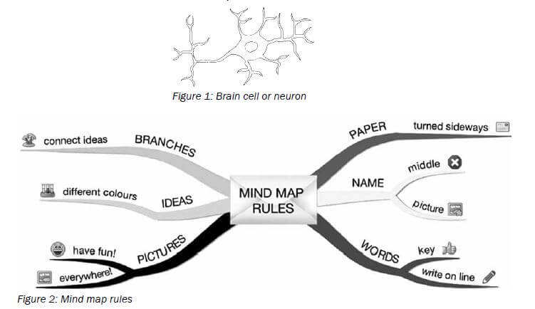
Mind maps work because they show information that we have to learn in the same way that our brains ‘see’ information.
As you study the mind maps in the guide, add pictures to each of the branches to help you remember the content.
You can make your own mind maps as you finish each section.
How to make your own mind maps:
- Turn your paper sideways so your brain has space to spread out in all directions.
- Decide on a name for your mind map that summarises the information you are going to put on it.
- Write the name in the middle and draw a circle, bubble or picture around it.
- Write only key words on your branches, not whole sentences. Keep it short and simple.
- Each branch should show a different idea. Use a different colour for each idea. Connect the information that belongs together. This will help build your understanding of the learning areas.
- Have fun adding pictures wherever you can. It does not matter if you can’t draw well.
On the day of the exam …
- Make sure you have all the necessary stationery for your exam, i.e. pens, pencils, eraser, protractor, compass, calculator (with new batteries). Make sure you bring your ID document and examination admission letter.
- Arrive on time, at least one hour before the start of the exam.
- Go to the toilet before entering the exam room. You don’t want to waste valuable time going to the toilet during the exam.
- Use the 10 minutes reading time to read the instructions carefully. This helps to ‘open’ the information in your brain. Start with the question you think is the easiest to get the flow going.
- Break the questions down to make sure you understand what is being asked. If you don’t answer the question properly you won’t get any marks for it. Look for the key words in the question to know how to answer it. A list of these words is on page xiv of this study guide.
- Try all the questions. Each question has some easy marks in it so make sure that you do all the questions in the exam.
- Never panic, even if the question seems difficult at first. It will be linked with something you have covered. Find the connection.
- Manage your time properly. Don’t waste time on questions you are unsure of. Move on and come back if time allows. You have 150 minutes (2½ hours) to answer each of the 150-mark Life Sciences question papers. Spend the following amounts of time on each question:
- Question 1: 50 marks = 45 minutes
- Question 2: 40 marks = 35 minutes
- Question 3: 40 marks = 35 minutes
- Question 4: 20 marks = 15 minutes
The remaining 20 minutes can be used to check your answers and attempt to answer any question that you might have left out.
- Check weighting - how many marks have been allocated for your answer? Take note of the ticks in this study guide as examples of marks allocated. Do not give more or less information than is required.
- Write big and bold and clearly. You will get more marks if the marker can read your answer clearly.
Question words to help you answer questions
It is important to look for the question words (the words that tell you what to do) to correctly understand what the examiner is asking. Use the words in the table below as a guide when answering questions.
Question word | What is required of you |
Analyse | Separate, examine and interpret |
Calculate | This means a numerical answer is required - in general, you should show your working, especially where two or more steps are involved |
Classify | Group things based on common characteristics |
Compare | Point out or show both similarities and differences between things, concepts or phenomena |
Define | Give a clear meaning |
Describe | State in words (using diagrams where appropriate) the main points of a structure/process/phenomenon/investigation |
Determine | To calculate something, or to discover the answer by examining evidence |
Differentiate | Use differences to qualify categories |
Discuss | Consider all information and reach a conclusion |
Explain | Make clear; interpret and spell out |
Identify | Name the essential characteristics |
Label | Identify on a diagram or drawing |
List | Write a list of items, with no additional detail |
Mention | Refer to relevant points |
Name | Give the name (proper noun) of something |
State | Write down information without discussion |
Suggest | Offer an explanation or a solution |
Tabulate | Draw a table and indicate the answers as direct pairs |

Learner’s checklist
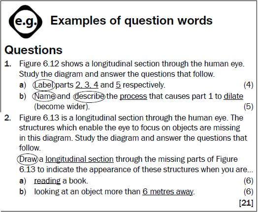
Use this checklist to monitor your progress when preparing for the examination.
The ticks (✔) tell you which aspects of the curriculum are covered in this study guide. The stars (*) tell you to go to textbooks and class notes.
TOPIC | ASPECT | Covered in The Study Guide | I do not understand | I understand |
DNA: The code of life | Discovery of DNA structure | * | ||
Nucleic acids terminology | ✔ | |||
Structure of DNA and RNA | ✔ | |||
Differences between DNA and RNA | ✔ | |||
DNA replication and the importance thereof | ✔ | |||
DNA profiling | ✔ | |||
Protein synthesis | ✔ | |||
Meiosis | The process of meiosis using diagrams | ✔ | ||
Significance of meiosis | ✔ | |||
Abnormal meiosis | ✔ | |||
Differences between meiosis I and meiosis II | ✔ | |||
Reproduction in vertebrates | Diversity in reproductive strategies | ✔ | ||
Human reproduction | Male reproductive system | ✔ | ||
Female reproductive system | ✔ | |||
Puberty | ✔ | |||
Menstrual cycle | ✔ | |||
Development of the foetus | ✔ | |||
Gestation | ✔ | |||
Role of placenta | ✔ | |||
Genetics and inheritance | Genetic terminology | ✔ | ||
Complete dominance | ✔ | |||
Incomplete dominance | ✔ | |||
Co-dominance | ✔ | |||
Inheritance of sex | ✔ | |||
Sex-linked characteristics | ✔ | |||
Dihybrid crossing | ✔ | |||
Mutations | ✔ | |||
Pedigree diagrams | ✔ | |||
Genetic engineering | ✔ | |||
Genetic counselling | ✔ | |||
Paternity testing and DNA fingerprinting | ✔ | |||
Responding to the environment: Humans | The brain | ✔ | ||
Neurons, reflex actions and reflex arcs | ✔ | |||
Peripheral nervous system | * | |||
Autonomic nervous system | * | |||
Brain disorders and injuries | * | |||
Effects of drugs on the central nervous system | * | |||
Structure and functions of parts of the eye | ✔ |
| Covered in The Study Guide | I do not understand | I understand | ||
Responding to the environment: Humans | Accommodation | ✔ | ||
Pupillary mechanism | ✔ | |||
Visual defects | ✔ | |||
Structure and functions of parts of the ear | ✔ | |||
Hearing | ✔ | |||
Balance | ✔ | |||
Hearing defects | ✔ | |||
Human endocrine system | Glands and the hormones they secrete | ✔ | ||
Negative feedback - glucose | ✔ | |||
Negative feedback - thyroxin | ✔ | |||
Homeostasis in humans | Negative feedback - glucose | ✔ | ||
Negative feedback - carbon dioxide | ✔ | |||
Negative feedback - water | ✔ | |||
Negative feedback - salts | ✔ | |||
The role of the skin on hot and cold days | ✔ | |||
Responding to the environment: Plants | Functions of auxins, gibberillins and abscisic acid | ✔ | ||
Role of auxins in phototropism and geotropism | ✔ | |||
Plant defence mechanisms is included | ✔ | |||
Evolution | Evidence for evolution | ✔ | ||
Sources of variation | ✔ | |||
Lamarck and Darwin’s theories | ✔ | |||
Natural and artificial selection | ✔ | |||
Punctuated equilibrium | ✔ | |||
Speciation | ✔ | |||
Mechanisms for reproductive isolation | ✔ | |||
Evolution in present times | ✔ | |||
Human evolution: similarities to African apes | ✔ | |||
Human evolution: differences from African apes | ✔ | |||
Major phases in hominid evolution | ✔ | |||
Out of Africa hypothesis | ✔ | |||
Phylogenetic trees | ✔ | |||
Human impact on the environment | Atmosphere and climate change | ✔ | ||
Water availability | ✔ | |||
Water quality | ✔ | |||
Food security | ✔ | |||
Loss of biodiversity | ✔ | |||
Waste disposal | ✔ | |||
Skills | Draw a line graph | ✔ | ||
Draw a bar graph | ✔ | |||
Draw a histogram | ✔ | |||
Draw a pie chart | ✔ | |||
Answering essay questions | ✔ |