Animal Reproduction - Agricultural Sciences Grade 12 Study Guides and Notes
Share via Whatsapp Join our WhatsApp Group Join our Telegram Group- Overview
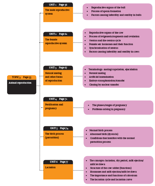
- The male reproductive system
- The female reproductive system
- Natural mating and other forms of reproduction
- Fertilisation and pregnancy
- The birth process(parturition)
- Lactation
- Topic Questions
Overview
The male reproductive system
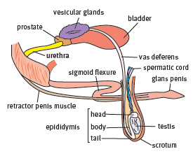
1. Reproductive organs of the bull
The reproductive organs of the bull can be divided into the:
- primary reproductive organs (mainly involved in reproduction) and
- the secondary reproductive organs (accessory or additional organs that assist with the production and delivery of semen).
Structure of primary male reproductive organs
Each primary male reproductive organ has a unique structure.
Structure of primary male reproductive organs
| Organ | Structure |
| Scrotum | Composed of skin on the outside and connective tissue and muscle on the the inside. It holds the two testes in place outside the body. The scrotum hangsdown between the hind legs of the bull |
| Testes | A bull has two testes which are contained in a bag of skin or scrotum. When thebull calf is born the testes are held inside the body cavity, but soon after birththe testes descend through the inguinal canal into the scrotum. The testes then hang inside the scrotum between the hind legs of the animal, suspended by aspermatic cord which consists of connective tissue and blood vessels.The testes are composed mostly of long, winding seminiferous tubuleswhich are about 1 km in length. The seminiferous tubules are surroundedby interstitial or supporting tissue. |
| Epididymis | An organ which lies next to the testes. It contains a long, convoluted tube calledthe ductus epididymis. The head of the epididymis lies against the upper partof the testes, the body lies over the surface of the testes and the tail on thelower edge of the testes. |
| Vas deferens | The tail of the epididymis passes into a tube called the vas deferens. The last 10 cm of the vas deferens is thicker than the start of the tube and it is known asthe ampulla. The vas deferens connects the epididymis to the urethra. |
| Penis | The male organ which lies outside the body and allows mating (copulation) totake place. Consists mainly of spongy tissue which becomes firm or erect whenthe blood vessels become filled.The root of the penis is attached internally to the pelvis. The body of the nonerectpenis lies in an S-shaped curve which is referred to as a sigmoid flexure.The tip is known as the glans penis. The penis at rest lies within a sheath ofskin called the prepuce. The penis is extruded through the preputial opening inthe sheath when bulls mate or urinate. |
| Urethra | A muscular tube extending from the bladder. It passes through the inside ofthe penis and opens at the glans penis. The vas deferens and the ducts of theaccessory sex glands also open into the urethra. |
 Structure of secondary male reproductive organs
The secondary or accessory sex glands of the bull are a group of glands behind the bladder in the pelvic cavity.
- They are in close contact with the ampulla of the vas deferens and the urethra.
- Vesicular glands (seminal vesicles) are
- the largest accessory glands
- they are paired
- consist of two vesicles or parts which are roughly 10 cm long
- have a knobbly surface.
- They lie above the bladder and the two vesicles form a V-shape around the ampulla of the vas deferens.
- Prostate gland surrounds the urethra where it leaves the bladder in the form of a ring.
- Cowper’s or bulbo-urethral glands are
- paired glands just behind the prostate
- on either side of the pelvic urethra.
Functions of the male reproductive
| Functions of primary male reproductive organs | |
| Organ | Structure |
| Scrotum | Main function is to protect the testes and regulate their temperature. The testes must be a few degrees cooler than body temperature to produce healthy sperm. This is why the testes are outside the body cavity. The scrotum helps to regulate the temperature in two ways:
|
| Testes | Function is to produce sperm or spermatozoa in the seminiferous tubules. The testes also produce the male sex hormone, testosterone. |
| Epididymis | Function is to concentrate, store and allow the maturation of the spermatozoa that originate from the testes. |
| Vas deferens | Function is to conduct sperm from the epididymis to the urethra during the process of ejaculation. |
| Penis | The male organ used for mating or copulation. It must become erect before the bull can mate. It becomes erect in response to the presence of a cow on heat, which causes the spongy tissue of the penis to fill with blood (called an erection). The penis lengthens and the sigmoid flexure straightens out. When the bull loses his erection the retractor muscle contracts and the penis returns to its original curved shape. |
| Urethra | Dual function:
|
Function of accessory sex glands
The accessory glands produce secretions or fluids which help spermatozoa to survive and travel from the testis to the urethra of the penis during ejaculation.
When the spermatozoa are suspended in the secretions of the accessory glands, the fluid is referred to as semen.
- Vesicular glands
- secrete a sticky, yellowish fluid which makes up 50% of the volume of semen in the bull
- secretion is needed to feed the spermatozoa, and correct the pH and osmotic pressure of the seminal fluid.
- Prostate gland
- secretes a watery, alkaline fluid that keeps the pH constant for the spermatozoa.
- lubricates and cleans the urethra which assists the movement of sperm during ejaculation.
- Cowper’s or bulbo-urethral glands
- produce an alkaline fluid which has the same function as the prostate gland.
2. Process of sperm formation
The male cell of reproduction is the spermatozoan or sperm cell. It is a microscopically small body that appears to have a head, neck and tail. The process of spermatozoa formation is called spermatogenesis. There are four stages in the process:
- Division of the spermatogonia (mitosis)
- Spermatogonia are cells in the wall of the seminiferous tubules of the testes.
- These cells divide by mitotic division and produce primary spermatocytes.
- Division of the primary spermatocytes (first meiosis)
- A primary spermatocyte undergoes meiosis to produce two secondary haploid spermatocytes.
- Division of the secondary spermatocytes (second meiosis)
- Each secondary spermatocyte produces two spermatids.
- Development of sperm cells
- The spermatids now develop into sperm cells (spermatozoa)
- consists of a head, neck and tail
- DNA is contained in the head of the spermatozoa.
3. Factors causing infertility and sterility in bulls
In some cases infertility may be permanent, in which case the bull is called sterile.
Lack of libido as a cause of infertility: Healthy adult bulls may show lack of libido or sexual urge due to the following reasons.
- Immaturity:
- Young bulls that have not reached full puberty may have low or no libido.
Onset of puberty varies with breed but is usually at 12–15 months of age. - Inexperience:
- Young and inexperienced bulls may have poor erections. They will require some time to practice mating.
- Overwork:
- Bulls that have to walk far to search for females over a wide area can often be exhausted from walking and lack of food or water. They will recover if rested and cared for properly.
- Disease:
- Various diseases which cause pain or loss of condition will reduce the bull’s libido.
- Temperament:
- Poor handling and change of environment may stress the bull and reduce his libido.
- Breed differences:
- European breeds mount animals frequently while Zebu animals will only mount females in oestrus.
- Old age:
- When bulls become too old they may show a lack of libido as a result of reduced testosterone secretion.
- Incorrect feeding
- Bulls may have poor libido due to nutritional problems. Underfeeding will cause a lack of energy while overfeeding will cause the bull to become fat and lazy. Specific deficiencies like vitamin A or protein deficiencies may cause poor libido.
Inability to mate as a cause of infertility: Bulls may produce semen of good quality but be unable to mount the cow or mate successfully. Reasons include:
- Conformation:
- Poorly developed hind legs or back muscles will affect the ability of the bull to mount the cow.
- Conditions of limbs:
- Foot or joint diseases can cause problems with mating because they prevent the bull from mounting.
- Injuries:
- An injury to the bull’s penis may prevent the bull from mating. This can be caused by thorns or barbed wire.
- Congenital abnormalities:
- A small number of bulls may be born with abnormalities of the genitalia which prevent them mating successfully.
Inability to fertilise as a cause of sterility: This happens when a bull shows good libido and is able to mate normally but fails to fertilise cows. Reasons include:
- Climate:
- Bulls that are not adapted to a hot climate may have a low sperm count.
- Diseases:
Various diseases (e.g. vibrio or campylobacteriosis and trichomoniasis) transmitted during mating may cause the failure of conception. - Diseases that cause a high fever (e.g. heartwater and redwater) may temporarily affect the sperm production of the bull. Lumpy skin disease (a viral infection) can cause permanent damage to the scrotum and testes. Some infections can cause orchitis (inflammation of the tissues of the testes).
- Malnutrition and exhaustion:
- These factors will cause a temporary reduction in sperm count which can be reversed if the bull is rested and fed correctly.
- Congenital defects:
- Inborn defects of the testes can make the bull sterile. e.g. producing abnormal sperm, scrotal hernia (testes become damaged) and cryptorchidism (testes fail to descend).
The female reproductive system
1. Reproductive organs of the cow
The reproductive organs of the cow are involved in the processes of ovulation, mating, fertilisation, pregnancy and calving. They are mostly located inside the abdominal cavity. They can be divided into two categories:
- the primary reproductive organs and
- the secondary reproductive organs.
Structure of female primary reproductive organ
Primary reproductive organ of the cow
The ovary is the primary reproductive organ of the cow. There are two ovaries, one on either side of the abdomen.
- They are attached by the fallopian tubes to the point of the horn of the uterus.
- They contain the ova or eggs of the cow.
When the heifer is born her ovary contains 30–400 000 unripe haploid ova or primary oocytes. When the cow reaches puberty a few ova will ripen every 21 days.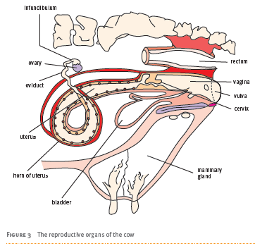
Structure of female secondary reproductive
Structure of secondary female reproductive
| Organ | Structure |
| Fallopian tubes | These tubes are also called the oviducts and they are two winding tubes which pass from the horns of the uterus to the ovaries. Each tube forms a funnel shaped body at the ovary called the infundibulum.the horns of the uterus to the ovaries. Each tube forms a funnel shaped body at the ovary called the infundibulum. |
| Uterus | A muscular tube which can be divided into various parts: the thick neck or cervix, the body and the horns. The uterus is lined on the inside with a layer called the endometrium. |
| Vagina | A muscular tube. It is adjacent to the cervix of the uterus on one end and it opens to the outside through the vulva or lips of the vagina on the other end. The hymen is the membrane at the entrance of the vagina. |
| Udder | Also known as the mammary gland, it is situated outside the body cavity of the cow. |
Functions of the female reproductive organs
Function of female reproductive organs
| Organ | Organ Structure |
| Ovaries | Produce the female sex cells called the ova or eggs which are then released into the funnel of the fallopian tubes during the process of ovulation. The ova develop inside a fluid-filled follicle (Graafian follicle). Oocytes develop every 21 days in response to the hormone FSH. |
| Fallopian tubes | They pick up the ovum which is released from the ovary during ovulation. Fine hairs or cilia help to move the ovum down the tube to the waiting sperm. |
| Uterus | Is usually tightly closed but during oestrus the wall relaxes and the cervix opens. The lining or endometrium contains glands which nourish the embryo before it implants. When the embryo implants, the blood supply of the endometrium nourishes it by being in close contact through the cotyledons that form on the foetal membrane. This forms the placenta in cattle. |
| Vagina | Has a number of functions. It receives the penis of the bull during mating and the semen released during mating. The semen travels up the vagina and through the cervix and the uterus. It then enters the fallopian tubes to effect fertilisation. The vagina is also the birth canal through which the foetus passes into the outside world. |
| Udder | Produces milk to feed the newborn animal. |
2. Process of ovigenesis/oogenesis and ovulation
The process of development of the egg (female sex cells or ova) in a cow is called ovigenesis or oogenesis.
- At birth, a heifer’s ovary contains thousands of primary oocytes or follicles.
- When she reaches puberty, under the influence of a number of hormones, a few primary follicles begin to develop. They contain a primary oocyte or egg cell which is surrounded by a layer of cells.
- These cells multiply and form a fluid-filled cavity in which the oocyte is suspended (Graafian follicle).
- When the oocyte becomes a mature ovum, the follicle ruptures and the ripe ovum is released into the fallopian tube which is called ovulation.
3. Oestrus and the oestrus cycle
Oestrus is the term used to describe the readiness of the cow for mating. It occurs at the stage in the 21-day oestrus cycle at which the cow ovulates (in heat) and is therefore ready to be fertilised by the bull.
Oestrus cycle
The oestrus cycle is a 21-day period during which a follicle (or follicles) develops into a mature ovum which is then released during the process of ovulation. The cycle is controlled by various hormones. A number of different processes take place during the oestrus cycle, which is divided into four phases.
Phase 1: Oestrus
- Days 0–1:
- The cow is in oestrus (on heat) for about 18 hours. The mature follicle usually ruptures 12 hours later.
Phase 2: Metoestrus
- Days 1–2:
- The cells of the ruptured follicle develop into a corpus luteum (CL).
- Days 2–5:
- The CL grows rapidly and developing follicles regress.
Phase 3: Dioestrus
- Days 5–16:
- The CL develops to its maximum size and function and produces the hormone progesterone. This hormone suppresses the formation of other follicles.
Phase 4: Proestrus
- Days 16–18:
- If fertilisation has not yet taken place, the uterus secretes the substance prostaglandin which signals the CL to regress rapidly.
- Days 18–20:
- Primary follicles begin to grow again and release oestrogen.
- Day 21:
- The oestrogen level rises and causes the onset of heat. The progesterone level decreases, the mature follicle ruptures and ovulation occurs.
However, if fertilisation occurs then the CL continues to produce progesterone:
- no follicles mature
- no ovulation takes place
- the cow will not return to heat again.
The progesterone produced then prepares the uterus for the implantation of the foetus.
External signs of oestrus
Cows show certain external signs and changes in behaviour under the influence of the hormone oestrogen, which show that she is ready for mating.
Behavioural changes
- The cow becomes excitable and begins to sniff, ride or mount other cows. A cow that has been ridden will show soiling with dung or mud around the hip bones. Cows in oestrus often stop eating and their milk production drops.
They show an interest in the bull and will stand to be mounted when ready to be mated. This is called standing heat.
Anatomical changes
- The vulva becomes moist, red and slightly swollen.
- The cow may also secrete a bullstring – a clear mucous secretion that dangles from the vagina.
Aids for oestrus detection
- A chalk is applied to the chin and the tail heads and will indicate when the cow is being ridden by other cows or is riding other cows.
- Pressure detection devices like the Kamar device – a white adhesive strip which is placed between the tailhead and the hip bone. The strip changes colour from white to red when the cow has been ridden.
4. Female sex hormones and their function
Sex hormones secreted by endocrine glands control the oestrus cycle of the female.
Follicle-stimulating hormone (FSH)
- The pituitary gland found at the base of the brain secretes FSH which stimulates the follicles to develop. These follicles secrete oestrogen as they develop.
- When oestrogen reaches a certain level, FSH secretion is inhibited and the pituitary gland secretes luteinising hormone (LH).
Luteinising hormone (LH)
- This hormone is secreted by the pituitary gland in response to the maturation of the Graafian follicle. This causes the follicle to rupture and develop a CL.
Oestrogen
- Oestrogen is produced mainly by the developing egg or Graafian follicle.
- The hormone is responsible for the development of the secondary sexual organs at puberty and the oestrus cycle after puberty.
- The hormone causes the external signs of oestrus and the behavioural changes in the cow which cause her to seek out and accept the bull.
Progesterone
- Progesterone is produced by the developing CL and prepares the uterus lining for the fertilised ovum (embryo).
- If fertilisation occurs, the CL will remain on the ovary and continue to produce progesterone throughout the development of the foetus (pregnancy).
5. Synchronisation of oestrus
Synchronisation of oestrus is when all the cows are on heat (oestrus) at the same time.
Techniques of synchronisation of oestrus (heat)
There are various techniques, involving the administration of different hormones or substances which affect the oestrus cycle, to synchronise oestrus.
Prostaglandin injections
- Injection of this hormone in a herd of cows causes a regression of the CL.
Stimulates the cow’s own FSH which begins the maturation of the follicles on the ovary. Ensures that all of the cows in the herd will come into heat at more or less the same time.
Gonadotropin releasing hormone (GnRH) injections
- Causes new follicles to develop. Results in the appearance of oestrus in the group. Can be injected into cows at various stages of the oestrus cycle.
Progestin or progestogen administration
- Synthetic forms of progesterone are used as vaginal implants or they are given in feed. The release of progesterone into the cow’s system at a constant rate prolongs the luteal phase. When the implant is removed or the feeding is stopped, the CL regresses and oestrus begins 36–60 hours later.
Advantages and disadvantages of synchronisation of oestrus
Table 1 The advantages and disadvantages of synchronisation of oestrus
| Advantages of synchronisation of oestrus | Disadvantages of synchronisation of oestrus |
|
|
6. Factors causing infertility and sterility in cows
Congenital or inborn factors include:
- underdevelopment of the reproductive organs, defects of the reproductive organs, freemartinism, prolapse or dropping out of the vagina, and persistence of the hymen which prevents sperm from entering the reproductive tract.
Injuries to the reproductive tract during mating, birth or the passing of the placenta can cause adhesions or blockages in the reproductive tract.
- This may prevent fertilisation.
Ovarian problems may cause a number of reproductive difficulties.
- These problems include failure to come on heat (anoestrus) and failure to ovulate.
Some of the causes of ovarian problems are disease, poor nutrition and very hot temperatures.
Diseases can cause infertility in cows.
- These include specific diseases of the reproductive tract like brucellosis, trichomoniasis and vibriosis (campylobacteriosis), general diseases which affect the overall health of the animal and non-specific infections such as those that result from tissue damage during difficult calving.
Malnutrition can affect the fertility of the cow. For example:
- underfeeding will cause a delay in puberty, while overweight cows will also show fertility problems.
Management must be handled correctly. For example:
- cows that are mated at the wrong time, subjected to unhygienic conditions and poorly adapted to their environment may develop fertility problems.
Factors causing infertility in cows
Cows are infertile when they are neither normally fertile nor completely sterile. So, infertility is thus not necessarily permanent, and can be caused by factors such as injuries to the reproductive tract, ovarian problems, diseases, malnutrition and poor livestock management.
Factors causing sterility in cows
Cows are deemed sterile when they have the physiological inability to effect sexual reproduction. In other words, sterility is a total loss of fertility. Sterility is thus caused when the factors that result in infertility persist. These can be:
- diseases of the genital organs (e.g. retained placenta)
- infections (e.g. trichomoniasis)
- physiological (e.g. repeat breeding)
- anatomical (e.g. freemartinism).
Natural mating and other forms of reproduction
1. Mating or copulation and ejaculation
Mating or copulation is when the male inseminates the female by penetrating the female genital tract and ejaculating sperm from the epididymis of the testis.
Natural mating
The normal method and most important form of insemination in farm animals is natural mating. This is when the bull identifies a cow on heat and mates with her.
- The bull is stimulated to mate by the presence of the oestrus cow.
- The drive to mate is regulated by his libido, which is under hormonal control.
The five main stages of mating or copulation
- Sexual display/courtship behaviour/pattern
- The bull is attracted to a cow on heat by her behaviour and shows an interest in her. He may sniff her hindquarters and curl his lip in a flehmen reaction. He may also rest his head on her rump.
Erection
- The penis of the bull becomes erect when he identifies a cow on heat. The erection occurs due to the dilation of blood vessels in the spongy tissue. Then the accessory glands begin to discharge their secretions into the urethra to lubricate and protect the sperm during ejaculation.
Mounting
- The cow will stand still if she is ready to be mated and allow the bull to jump, raise his forequarters and straddle her back. He is able to place the penis near to the vulva in this position.
Penetration
- The bull drives his erect penis into the vagina of the cow when he is in the correct position.
Ejaculation
- The sperm is now discharged from the epididymis. It travels down the vas deferens, enters the urethra and is discharged through the tip of the penis into the vagina. The bull dismounts from the cow once he has ejaculated.
Artificial insemination (AI)
Artificial insemination (AI) in cattle was originally developed to prevent the infection of cows with venereal diseases such as vibriosis.
Later scientists realised that this method was a very quick way to improve the quality of herds by using semen from bulls that had superior genetic characteristics
- One good bull can be used to inseminate many more cows than it is able to serve
- Today AI is used for genetic improvement in dairy and beef herds.
- In AI semen is collected from the male animal under controlled conditions and then it is artificially inserted (inseminating) into the reproductive tract of the female.
Basic requirements for successul AI
Proper collection and storage of semen:
- Semen is collected by leading a bull to a ‘cow’ (a dummy) or another bull (the teaser bull).
- When the bull mounts the ‘cow’ or dummy, an artificial vagina (AV) is placed over the bull’s penis to collect the ejaculate.
- Some operators use electrical stimulation or an electro-ejaculator to cause ejaculation.
Good quality semen:
- Good quality semen contains a large number of spermatozoa that move around rapidly and a low percentage of abnormalities such as loose tails or double heads.
Good technique (insemination):
- The plastic straw is taken out of the liquid nitrogen and the thawed semen is inserted into a special pipette.
- The operator then places an arm in the cow’s rectum, grasps the cervix and pushes it downwards gently. The operator uses the other hand to insert the pipette through the vulva and then through the cervix
- Half of the semen is deposited (as above) and the rest is deposited into the uterine horn.
- The pipette is then withdrawn and the cervix is then massaged → This causes the secretion of oxytocin, which causes the uterus to contract. This action forces the sperm forward in the reproductive tract.
Accurate timing (detecting oestrus in cow): Beef cows need to be inseminated as soon as they show signs of heat, whereas dairy cows should be inseminated 12 hours after the detection of heat.
Types of semen diluent and their function Semen collected from the animal must be suspended in a liquid diluent. This diluent has various components and serves a number of functions. Semen diluent is usually made up with equal parts of a buffer and a protectant.
One of the main functions of diluents is to extend the semen so that it can be used to inseminate more animals. Diluents also have buffering functions (stabilises the pH) and protect the spermatozoa from being damaged by the freezing process which occurs when the semen is stored in liquid nitrogen.
The advantages and disadvantages of AI
| Comparing the advantages and disadvantages of AI | |
| Advantages of AI | Disadvantages of AI |
| Rapid genetic improvement of a herd is possible | Rapid spread of disease if semen is infected |
| Venereal diseases can be controlled | Need very good hygienic practice and management |
| Cheaper than buying and keeping expensive stud bulls (± one bull per 30–60 cows) | Rapid spread of undesirable genetic material |
| Breeding programme has greater flexibility because of wide choice of bull semen | Narrowing of genetic base (reduced genetic diversity) |
| Oestrus synchronisation and AI make it possible to manage breeding better | |
| Individual good bulls can supply semen to large number of farms even after his death | |
Embryo transplantation/transfer (ET)
Embryo transplantation/transfer (ET) is the transfer of a large number of fertilised embryos from a good quality donor cow into the uterus of many recipient or surrogate cows. The aim is to allow a very good cow to produce a large number of offspring.
The first stage of ET is to administer FSH and cause superovulation in a chosen cow.
- The cow is then inseminated with good quality semen.
When the semen is fertilised, the embryos are harvested or removed from the donor cow by embryo flushing.
- This is done by inserting a tube into the uterus, adding sterile liquid and then sucking or washing (flushing) the embryos from the uterus.
The embryos are examined to ensure that they are healthy.
- Suitable embryos are then either frozen or implanted into recipient cows.
Finally the embryos grow in the recipients who give birth to the calves.
Advantages and disadvantages of ET
| Advantages of ET | Disadvantages of ET |
|
|
Cloning by nuclear transfer
Cloning in biotechnology refers to processes used to create copies of DNA fragments (molecular cloning), cells (cell cloning) or organisms. Cloning by nuclear transfer involves producing an exact copy of an existing animal. It is done by injecting the nucleus of a somatic cell from the animal to be cloned into an unfertilised ovum or egg cell. The ovum’s own DNA is removed. This results in a new cell that will divide normally, forming an embryo that is identical to the donor animal. The embryo is placed in the uterus of a surrogate mother where it grows to term.
- Types and aims of cloning:
- Reproductive cloning
- Farm animals are cloned to reproduce individuals with valuable genetic material (e.g. farmers may want to reproduce animals that have a particular advantage such as disease resistance or exceptional production characteristics).
- Therapeutic cloning
- The main aim is to use embryonic stem cells, which have the unique ability to generate virtually all types of cells in an organism, to grow tissues in the laboratory that can be used to grow healthy tissue to replace injured or diseased tissues.
- It may also be possible to learn more about the molecular causes of disease by studying embryonic stem cell lines from cloned embryos derived from the cells of animals or humans with different diseases.
Types of cloning processes
| Reproductive cloning | Therapeutic cloning |
|
|
Advantages and disadvantages of cloning
| Advantages of cloning | Disadvantages of cloning |
|
|
Fertilisation and pregnancy
1. Reproduction terminology
Fertilisation:
- In animals, this involves the fusion of an ovum with a sperm, which eventually leads to the development of an embryo. It starts with copulation.
- After a male ejaculates, a large number of sperm cells move to the upper vagina (via contractions from the vagina) through the cervix and across the length of the uterus to meet the ovum.
- In cases where fertilisation occurs, the female usually ovulates during a period that extends from hours before copulation to a few days after.
Pregnancy:
- In animals, this is the period of reproduction during which a female carries one or more live offspring in the uterus through gestation.
- It begins when a fertilised zygote implants in the female’s uterus and ends once it leaves the uterus.
Placenta:
- This is a flattened circular organ in the uterus of pregnant mammals.
- It consists of vascular tissue in which oxygen and nutrients can pass from the mother’s blood into that of the foetus, and waste products can pass in the reverse direction.
- The placenta is expelled from the uterus at the birth of the foetus.
Freemartin:
- An imperfect sterile female calf that is the twin of a male calf whose hormones affected its development.
- Freemartinism occurs occasionally in twins which are male and female.
- It results from the fusion of placentas of the two foetuses.
- This causes the female foetus to be exposed to testosterone, resulting in a masculinised and infertile animal.
2. Fertilisation process
After mating or AI, millions of spermatozoa use the lashing movements of their tails to swim very rapidly up the reproductive tract to the fallopian tube.
- They are assisted by the contractions of the uterus.
- They wait for the ovum to arrive when they reach the fallopian tube.
- Once the ovum is released from the ovary, it is picked up by the fallopian tube and moved down the fallopian tube where the spermatozoa are waiting.
- At this stage many of the spermatozoa that reached the fallopian tube will have died off.
- These sperm release an enzyme called hyaluronidase which breaks down the outer layer of the ovum in a process called capacitation.
→It allows a single sperm to finally enter the ovum and join its DNA with that of the ovum which is called fertilisation.
→ The fertilised cell is called a zygote. It starts to divide and it is has different names at various stages of division (morula, blastocyst and finally embryo).
Multiple births, twinning and freemartins
Formation of multiple births (twins)
- If more than one ovum is shed during ovulation, then more than one zygote can be formed. For example, pigs normally have 6–14 piglets and sheep can give birth to twins or triplets. Multiple births are less common in cows but does occur.
The formation of the twins is dizygotic if two ova are fertilised and monozygotic when the embryo splits into two during development.
Formation of freemartins
- Dizygotic twins of different sexes occasionally develop an interconnected blood supply. If this happens, the heifer receives testosterone which causes the underdevelopment of the female sex organs and the formation of tiny ovaries. This results in a sterile freemartin. These heifers can be identified by external signs such as excess hair on the vulva and an underdeveloped udder and teats.
3. Phases or stages of pregnancy
The embryo remains in the fallopian tube for 4–5 days. It then moves down into the uterus where it lies loose in the uterus for the first month.
- The embryo then differentiates into three different layers, namely the endoderm, mesoderm and ectoderm which give rise to the various tissues and organs.
- Between the first and fourth months of pregnancy, the foetus attaches to the uterus and is enclosed in a number of membranes or layers.
- The amnion membrane surrounds the foetus directly and contains amniotic fluid
- The chorion, which is attached to the umbilicus of the foetus like the amnion, lies against the uterus wall and is filled with fluid.
- A small sac (allantois) gradually enlarges and becomes attached to the chorion.
- The embryo excretes urinary waste through the umbilicus into the allantoic sac.
- The fused chorio-allantois forms round areas of attachment to the uterine wall called cotyledons.
- In cows, cotyledons represent the placenta, or the area in which the blood supply of the foetus and the mother are closely in contact. This allows the embryo to receive oxygen and nutrients by diffusion and osmosis.
The phase/stage of the foetus The stage of the foetus begins 45 days after fertilisation. The foetus is clearly recognisable since all of its tissues are fully differentiated. During this stage the foetus grows in size, its organs develop further, tiny bones begin to calcify and hair begins to grow.
4. Problems arising in pregnancy
Resorption
- Occurs when a young embryo dies off before it implants and is partially or completely re-absorbed. Resorption is the most common cause of failed pregnancies. No external signs will be seen with resorption because the small and soft tissues are absorbed by the cow’s body.
- The reasons for resorption
- genetic problems which cause faulty development
- infections of the reproductive tract
- the breakdown of the corpus luteum which results in decreased progesterone levels and a deterioration of the uterine lining.
Abortions
- This is when the foetus dies and is passed out through the vagina. External signs of an abortion include if the foetus is found or the placental membranes are seen hanging from the vulva of the cow. The foetus may also remain in the uterus after infection (in the form of macerated, decomposed or mummified material). The soft tissues will disappear and leave a dry, hard foetus.
- The reasons for abortions
- Infections: Most common cause of an abortion. An infection often damages the organs or causes them to be malformed which results in death of the foetus. In some cases it causes placentitis or inflammation of the cotyledons or placenta which disrupts the mother’s blood supply.
- Genetic factors: A genetic defect to a vital organ may cause the foetus to die off and be aborted.
- Environmental factors: Malnutrition or rough handling will injure a pregnant cow and may cause the foetus to be aborted.
- Toxins: Various toxins, such as those found in poisonous plants, may cause the cow to abort.
- Hormonal disturbances: A breakdown of the corpus luteum during pregnancy will lead to a decrease in the progesterone levels and cause the cow to abort.
The birth process (parturition)
1. Parturition (birth)
Physical signs show that the cow is approaching parturition:
- abdomen: becomes enlarged during the foetal stages and appears to drop due to the relaxation of the ligaments of the pelvic canal
- udder: begins to develop in the last few weeks of pregnancy and shows signs of producing milk a few days before calving
- vulva: begins to swell and discharge a thick mucous
- behaviour: on the day of calving the cow stops eating, isolates herself, searches for a place to give birth, shows restlessness and discomfort, and attempts to urinate often.
Functions of the membranes covering the foetus
| Foetal membranes/layers and their functions | |
| Foetal membrane/layer | Function |
| amnion (inner membrane) | Encloses amniotic fluid which serves as shock absorber around foetus and provides lubrication for birth of calf |
| chorion | Fuses with allantois |
| allantois (outer membrane) | Encloses the allantoic fluid which collects the excretory products from the foetus |
| chorio-allantois | Attaches to uterus wall and forms the cotyledons of the placenta which allows exchange of nutrients between the foetus and mother |
Stages/phases of parturition
Stage 1: Preparatory
- The uterus begins to contract involuntarily at intervals during the preparatory stage. The cervix opens as a result of hormone secretion and is pushed open further by the fluid-filled foetal membranes that press against the uterine end of the cervix. The allantois or outer membrane ruptures and releases allantoic fluid which lubricates the birth canal.
Stage 2: Expulsion of the foetus
- The cow usually lies down in this stage. In addition to involuntary uterine contractions, the cow begins to strain using her abdominal muscles to expel the foetus from the birth canal. These combined contractions propel the foetus through the dilated cervix. The amnion (inner membrane) ruptures which releases fluid to help lubricate the passage of the foetus. The foetus is expelled and the umbilicus pulls loose from the uterine wall. The calf’s breathing reflex is stimulated because it no longer receives oxygen through the umbilicus or navel.
Stage 3: Expulsion of the afterbirth
- A period of rest follows the expulsion of the foetus from the uterus. The uterine muscles then contract again and expel the so-called afterbirth, which is composed of the placenta, umbilical cord and foetal membranes.
2. Conditions that interfere with normal parturition
Dystocia
Signs of dystocia (birth problems) include the bursting of the allantois, straining by the cow or the appearance of the feet or head followed by no further activity.
Causes of dystocia
The causes of dystocia can be maternal (due to the cow) or of foetal origin.
Causes of dystocia
| Foetal causes | Maternal causes |
| Abnormal presentation Twin pregnancy Over large calves Dead calves Deformed calves | Uterine torsion Metabolic diseases Over fat cows Anatomical defects of pelvis Injuries |
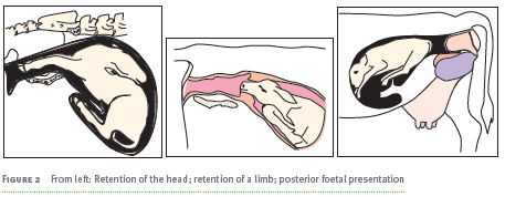
Correct birth positions of a calf in the uterus
There are two normal birth positions. In the anterior presentation, the head and feet come out first.
Posterior presentation is when the hind legs come out first, followed by the rest of the body.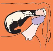
Other conditions that can interfere with normal parturition
Vaginal or vulval tears
- Vaginal or vulval tears usually result from the use of force to remove a calf. The most common causes are not using lubrication, allowing the hooves of the calf to penetrate the vagina or the use of extreme force.
Prolapsed uterus
- The uterus may come out of the vagina after the birth process under certain conditions, for example when the cow has to strain very hard during labour.
Calving paralysis
- If a calf is pressed against the cow’s pelvic cavity, a nerve will be pinched between the pelvic cavity and the bulk of the calf. This nerve damage can result in paralysis. The cow will be able to stand but will not be able to use the hind leg on one side.
3. Principal factors causing retention of the placenta in cows
The expulsion of the placenta usually takes place naturally when the uterus contracts some hours after the birth process. However, some direct and indirect factors can cause the placenta to remain inside the uterus where it may rot and cause health problems.
Table 3 Direct and indirect factors causing the retention of the placenta in cows
| Direct factors | Indirect factors |
|
|
Lactation
1. Structure and functions of the cow udder
The udder or mammary gland is the organ in which milk is produced. The udder of the cow consists of four parts or quarters which are completely separate from each other.
- The udder is covered with skin and is attached to the body by a number of ligaments. Each quarter of the udder is made up of a mass of secretory tissues or alveoli which are arranged in groups or lobules.
- The alveoli produce the components of milk. The milk produced in the alveoli moves into the small milk cavities and then moves down a series of ducts or tubes.
- The small milk ducts lead into the large milk ducts, and from there the milk travels downwards and Lactation, dry period, milk ejection pools in the gland cavity just above the teat canal. The entrance to the teat is
held closed by a ring of muscle. - The teat itself has a cavity and an opening which is called the teat orifice. The teat opening is held closed by a sphincter muscle which helps to prevent the entry of bacteria up the teat canal. Bacterial invasion of the udder causes an infection called mastitis which decreases milk production.
Lactation is the production of milk in order to nourish the newborn animal. Weaning occurs when the young animal is used to food other than its mother’s milk. In dairies, the calf is removed but the mother cow is milked daily.
This keeps up her milk production until she is dried off by the farmer. The dry period in dairy cows is the period after this drying off has occurred. The dry period usually occurs when the calf is weaned. In dairies the calf is removed so the farmer needs to imitate this by gradually reducing milking until lactation stops.
The milk ejection (milk let down) is the release of milk from the udder into the teats, which allows the calf to drink or the cow to be milked.
Composition of milk
Milk is a highly nutritious liquid which is specially designed for the growth and protection of young mammals. Each species produces milk with a slightly different composition. The milk from most species contains roughly 2–11% fat, 3–7% protein, 5% milk sugar, and vitamins and minerals. It is a very rich source of calcium which promotes bone growth in young animals.
Some of the milk components are obtained from the blood supply and these include water, minerals, blood proteins such as antibodies, vitamins and enzymes. Other components such as lactose (milk sugar), casein (milk protein) and fat (butterfat) are formed in the udder tissue itself. Milk production by the mammary gland tissue is controlled by the hormone somatotropin which is secreted late in pregnancy.
2. Hormones and milk ejection/milk let down
Two processes are important for the removal of milk from the udder: passive withdrawal and milk ejection or let down through the milk ejection reflex.
Passive withdrawal
Passive withdrawal of milk is the emptying of the larger ducts and cavities of the udder by neural stimulation of the skin and teats of the udder. This phase begins 5 to 10 seconds after stimulation of the udder. Between 40 and 50% of the milk can be removed passively from the udder.
Hormones involved in milk ejection/milk let down
For the remaining 50 to 60% milk to be removed, successful milk ejection has to take place. This happens due to the milk ejection reflex, which is a neuro-hormonal reflex.
- Milking or suckling stimulates nerve endings in the teats, bringing about the release of the hormone oxytocin from the pituitary gland 20 to 40 seconds later.
- Oxytocin causes the expulsion of milk from the alveoli by stimulating the epithelial cells of the ducts to push the milk down into the gland cavities and then into the teats.
- Oxytocin is present in the bloodstream for 6 to 7 minutes after stimulation, which therefore is the time available for milking a cow if the maximum yield is to be obtained from her.
- An excited or scared cow will secrete the hormone adrenalin. This hormone will inhibit milk ejection, which will prevent the cow from being milked properly.
Importance and functions of colostrum
The milk which is secreted just after the birth of the calf is called colostrum or first milk. It is very thick and provides nutrient rich food for the newborn as well as antibodies that are concentrated from the mother’s blood. The intestine of the calf is able to absorb the antibodies for the first 48 days of its life. The colostral antibodies are absorbed into the calf’s bloodstream instead of being broken down. They protect the calf against infectious diseases until the young animal develops its own. It is therefore very important that a newborn animal receives enough colostrum as soon as possible after birth and always within the first 48 hours.
3. The lactation cycle and lactation curve
The lactation cycle (period)
The dairy cow has been bred to supply a large quantity of milk for as long as possible. To achieve this, the cow must give birth to one calf a year.
- The lactation period begins after calving and three months later she should come on heat and be mated or inseminated.
- The cow is dried off in the last two months before she is due to calve again. This is known as the drying period.
- This means that her milk production is halted by reducing the milking of the udder.
- Drying off allows the udder two months to rest and recover before the cow calves again.
- The cow is therefore in lactation for roughly 300 days or 10 months.
- This 12 month cycle is called the lactation cycle. Farmers need to monitor the lactation cycle of their herd in order to manage them effectively. It is important to get cows pregnant as early as possible within the 12 month period.
- Dairy cows usually have 5–6 lactation cycles during their lifetime.
The lactation curve
The cow secretes colostrum for a few days just after calving and then she begins to produce normal milk.
- A good dairy cow produces roughly 40 litres per day at this early stage of lactation
- Milk production reaches a peak roughly 1–2 months after lactation begins.
- Thereafter the daily production gradually decreases until she is dried off two months before her next calf is due to be born.
- This fluctuation in the amount of milk produced is shown by a lactation curve.
- Dairy farmers record the lactation curve of the herd to monitor their production.
- This allows farmers to assess whether the cow’s milk production has reached its genetic potential.
- The amount of milk produced during the lactation cycle depends on genetics and the feeding of the animal.
Topic Questions
- Answer the questions below. Check your answers afterwards and do corrections.
- Give yourself one hour.
- Marks: 100
- Name the reproductive organ in which the following processes take place:
1.1 ovulation
1.2 fertilisation
1.3 spermatogenesis
1.4 implantation
1.5 lactation
1.6 parturition. (6) - Hormones are release during the oestrus cycle. Name a function of the following hormones in the oestrus cycle:
2.1 Progesterone
2.2 Oestrogen (2) - Outline the main difference between infertility and sterility. (2)
- List FIVE important factors to consider in the collection, examination and storage of semen to ensure that the product is of good quality. (5)
- It is important for a farmer to be able to detect oestrus in a cow.
5.1 Explain why it is important. (1)
5.2 Describe TWO signs of oestrus/heat in a cow. (4)
5.3 What is standing heat? (2)
5.4 When is the best time to mate/inseminate a cow? Distinguish between beef cattle and dairy cows. (2)
5.5 Is the oestrus cycle in cows seasonal? (1) - Describe the process of fertilisation. Explain where and how it takes place. (5)
- Can a monozygotic twin be a freemartin? Explain your answer. (3)
- Describe the difference between resorption and abortion. (5)
- Outline the sequence of events when a cow gives birth. List and briefly describe all of the important processes. (10)
- Incorrect foetal presentation is one of the main causes of dystocia in cows because it prevents the calf from moving through the birth canal.
10.1 What are the main incorrect foetal positions that cause problems? (3)
10.2 How are the positions referred to in (10.1) usually corrected? (1) - Milk production is under hormonal control.
11.1 Which hormone is involved in the control of lactation? (1)
11.2 Name FOUR common causes of a drop in milk production. (5)
11.3 What normal physiological process can cause a drop in milk production three months after calving? (1) - Name the main categories of causes for a lack of libido in bulls.
12.1 List four and provide a brief description of each one. (8) - Discuss the role of disease in the reproductive failure of cows. (6)
- Cloning is the process of producing an exact replica of an animal.
14.1 Would you use this technique as a replacement for natural mating? (2)
14.2 Give reasons for your answer. (3) - Discuss the importance of correct feeding during the reproductive process. Provide and elaborate on at least two factors. (6)
- Which THREE of these foetal presentations can cause dystocia?
16.1 Anterior presentation
16.2 Posterior presentation
16.3 Breech presentation
16.4 Anterior presentation with head retained
16.5 Posterior presentation with limb retained (3) - Name THREE conditions of the calf that can cause dystocia. (3)
- Name FOUR signs that indicate that a cow is about to calve. (4)
- Define milk let down. (1)
- Use the information below to draw a lactation curve. (5)
A good dairy cow produces roughly 40 litres per day at this early stage of lactation. Milk production reaches a peak roughly 1–2 months after lactation begins. Thereafter the daily production gradually decreases until she is dried off two months before her next calf is due to be born.