RESPONDING TO THE ENVIRONMENT- HUMANS GRADE 12 NOTES - LIFE SCIENCES STUDY GUIDES
Share via Whatsapp Join our WhatsApp Group Join our Telegram GroupRESPONDING TO THE ENVIRONMENT- HUMANS
LIFE SCIENCES
STUDY GUIDES AND NOTES
GRADE 12
CHAPTER 6: RESPONDING TO THE ENVIRONMENT - HUMANS
The human nervous system
The nervous system is responsible for processing and transmitting information throughout the body:
- It tells the body how to react to stimuli (changes in the environment to which the body responds). For example, it regulates body temperature on a hot or cold day. It is also responsible for the reflex action, for example, when you step on a pin or touch a hot surface.
- The nervous system also coordinates the various activities of the body, such as walking, hearing, seeing, and so on.
The central nervous system consists of the brain and the spinal cord.
6.1 The brain
6.1.1 Structure and functions of the brain
Figure 6.1 below shows the different parts of the brain and their functions.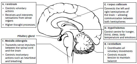 Figure 6.1 The structure and functions of the brain
Activity 1
Questions
Write down the name of the part which:
- Controls heartbeat (1)
- Contains the centres that control balance, muscle tone and equilibrium (1)
- Has centres that interpret what you see (1)
- Coordinates voluntary muscle movements (1)
- Controls body temperature (1) [5]
Answers to activity 1
- Medulla oblongata✔(1)
- Cerebellum✔ (1)
- Cerebrum✔ (1)
- Cerebellum✔ (1)
- Hypothalamus✔ (1) [5]
6.2 Neurons
Neurons are specialised cells which connect the brain and spinal cord to all other parts of the body.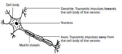 Figure 6.2 A neuron
There are three types of neurons, namely sensory (afferent) neurons, motor (efferent) neurons and interneurons (or connectors). Table 6.1 below shows the structure and function of these neurons.
| Type of neuron | Function | Structure |
| Sensory (afferent) neuron Senses the stimulus | Transmits impulses from the sense organs or receptors to the spinal cord and brain. | 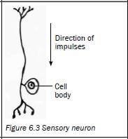 |
| Motor (efferent) neuron Response to the stimulus | Transmits impulses from the brain and spinal cord to the effectors (muscles and glands). The effectors bring about the response. | 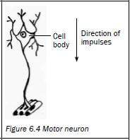 |
| Interneuron (connector) Found in the brain and the spinal cord. | Links the sensory neuron to the motor neuron | 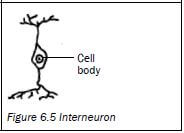 |
Table 6.1 Sensory, motor and interneurons
A synapse is the functional connection between the axon of one neuron, and the dendrites of another neuron.
6.3 Reflex arc
A reflex action is a quick, automatic action that involves the spinal cord and does not involve the brain. It is an important function to protect the body from harm. Examples are blinking the eye, coughing, sneezing, dilation and constriction of the pupil of the eye, and quickly withdrawing your hand when it touches a hot surface.
The reflex arc is the path along which an impulse is transmitted to bring about a response to a stimulus during a reflex action.
Figure 6.6 below shows what happens when you hold your finger close to a flame. The grey arrows represent the reflex arc.
The path of a reflex arc: |
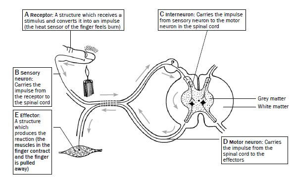
Figure 6.6 The reflex action of withdrawing a finger when placed in a flame
Activity 2
Questions
Use the diagram of the reflex arc in Figure 6.6 on page 44 to answer the following questions.
- Part B indicates the …
- dendrite of the motor neuron.
- axon of the motor neuron.
- dendrite of the sensory neuron.
- axon of the sensory neuron. (2)
- The correct sequence in which impulses move from the receptor to the effector in the reflex arc in Figure 6.6 is ...
- A → B → C → D→ E
- C → A → B → E→ D
- C → B → E → D→ A
- A → D → E → B→ C (2)
- Give the correct term for the following definitions:
- A structure which receives a stimulus and converts it into a impulse
- A structure which responds to a stimulus, e.g. a muscle or gland
- A neuron that carries impulses from the central nervous system to the effectors
- A neuron that carries impulses from the receptors to the central nervous system
- A neuron that carries impulses from a sensory neuron to a motor neuron in the spinal cord
- A very quick, automatic action that involves the spinal cord and not the brain
- The pathway along which an impulse is transmitted to bring about a response to a stimulus during a reflex action 7 × 1 = (7) [11]
Answers to activity 2
- C✔✔(2)
- A✔✔ (2)
-
- Receptor✔
- Effector✔
- Motor/efferent neuron✔
- Sensory/afferent neuron✔
- Interneuron✔/ connector
- Reflex action✔
- Reflex arc✔ 7 × 1 = (7) [11]
6.4 The human eye
Figure 6.7 below shows the different parts of the eye and their functions.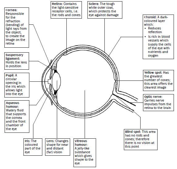
Figure 6.7 The structure of the eye
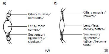
6.4.1 Accommodation
Accommodation is the adjustment of the shape of the lens to see objects clearly whether they are far away or close by. This is shown in Table 6.2 and Figures 6.8 and 6.9 below.
Distant vision (objects further than 6 m) | Near vision (objects closer than 6 m) |
1. Ciliary muscles relax | 1. Ciliary muscles contract |
2. Suspensory ligaments tighten (become taut) | 2. Suspensory ligaments slacken |
3. Tension on lens increases | 3. Tension on lens decreases |
4. Lens is less convex (flatter) | 4. Lens becomes more convex (more rounded) |
5. Light rays are refracted (bent) less | 5. Light rays are refracted (bent) more |
6. Light rays are focused onto the retina | 6. Light rays are focused onto the retina |
|
|
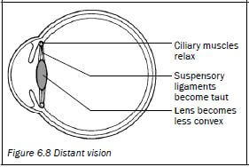
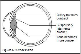
Table 6.2 Accommodation of the eye for distant and near vision
6.4.2 Pupillary mechanism
The pupillary mechanism (or pupil reflex) regulates the amount of light entering the eye by adjusting the size of the pupil. This is shown in Table 6.3 and Figures 6.10 and 6.11 below.
Light is bright | Light is dim |
1. Radial muscles of the iris relax | 1. Radial muscles of the iris contract |
2. Circular muscles of the iris contract | 2. Circular muscles of the iris relax |
3. Pupil constricts (gets smaller) | 3. Pupil widens (gets bigger) |
4. Less light enters the eye | 4. More light enters the eye |
|
|
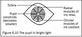
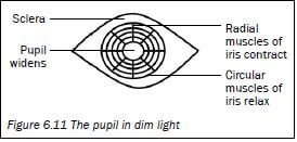
Table 6.3 Pupillary mechanism
6.4.3 Visual defects
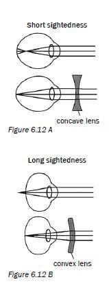 | Visual defect | Nature of the defect | Corrective measures |
| Short-sightedness Near objects can be seen clearly (myopia) |
| Wearing glasses with converging (biconcave) lensFigure 6.12 A | |
| Long-sightedness Objects far-away can be seen clearly (hyperopia) |
| Wearing glasses with converging (biconvex) lensFigure 6.12 B | |
| Astigmatism |
| Glasses with lenses shaped to correct the distortion | |
| Cataracts |
| Surgery to replace thelens with a synthetic lens |
Activity 3
Questions
- Figure 6.13 shows a longitudinal section through the human eye. Study the diagram and answer the questions that follow.
a) Label parts 2, 3, 4 and 5 respectively. (4)
b) Name and describe the process that causes part 1 to dilate (become wider). (5) - Figure 6.14 is a longitudinal section through the human eye. The structures which enable the eye to focus on objects are missing in this diagram. Study the diagram and answer the questions that follow.
Draw a longitudinal section through the missing parts of Figure 6.14 to indicate the appearance of these structures when you are ...
a) reading a book. (6)
b) looking at an object more than 6 metres away. (6) [21]
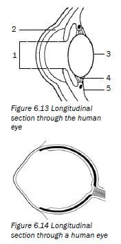
Answers to activity 3
-
a) 2 - Cornea✔
3 - Lens✔
4 - Suspensory ligaments✔
5 - Ciliary muscles✔/ body (4)
b) Pupillary mechanism✔/ pupil reflex
The radial muscles3of the iris contract3and the circular muscles✔relax.✔
The pupil dilates and more light enters the eye.✔ (5) -
 [21]
[21]
 [21]
[21]6.5 The human ear
6.5.1 Structure of the ear
The human ear consists of three main parts:
- The outer ear
- The middle ear
- The inner ear
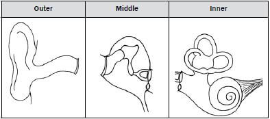
Figure 6.15 below shows the structure and function of each part of the human ear.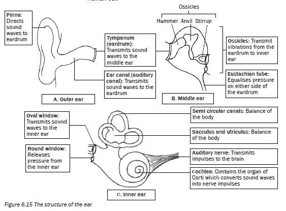
Figure 6.15 The structure of the ear
6.5.2 Hearing
Figure 6.16 below shows how the three parts of the ear work together to make it possible for us to hear. The grey arrows show the path of a sound wave.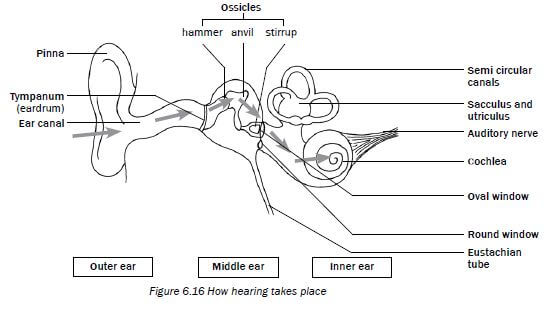
Figure 6.16 How hearing takes plac
Look at Figure 6.16 above and read the information in Table 6.4 below to understand how hearing takes place.
Part of ear | What it does during the hearing process |
Pinna | Traps the sound waves and directs them into the auditory canal. |
Tympanic membrane | Vibrates and transmits the vibrations to the ossicles in the middle ear. |
Ossicles | The ossicles amplify the vibrations and carry them via the middle ear to the membrane of the oval window. |
Oval window | Vibrates and causes pressure waves in the inner ear. |
Cochlea | These vibrations cause the sensory cells in the organ of Corti to be stimulated in the cochlea and nerve impulses are generated. |
Auditory nerve | Transmits nerve impulses to the cerebrum to be interpreted. |
Table 6.4 The hearing process
6.5.3 Balance
The human ear is responsible for balance in this way:
1. The cristae in the semicircular canals are stimulated by changes in the direction and speed of movement
2. The maculae in the sacculus and utriculus are stimulated by changes in the position of the head
When stimulated, the cristae and maculae convert the stimuli received into nerve impulses.
The nerve impulses are transported along the auditory nerve to the cerebellum to be interpreted.
The cerebellum then sends impulses to the muscles to restore balance.
6.5.4 Hearing defects
Hearing defect | Causes | Treatment |
Middle ear infection |
|
|
Deafness |
|
|
Table 6.5 Hearing defects
Activity 4
Questions
Study Figure 6.17 below and answer the questions that follow.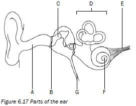
1. Identify the parts labelled B, C and F. (3)
2. Give the function of the pinna. (2)
3. Write the letter of the part which:
a) contains receptors for balance. (1)
b) equalises the pressure on either side of part B. (1)
c) transmits impulses to the brain. (1)
4. Describe how hearing occurs. (8) [16]
Answers to activity 4
1.
B -Tympanic membrane✔
C - Malleus/hammer/an ossicle✔
F - Cochlea✔(3)
2. It directs sound waves✔ into the auditory canal✔. (2)
3.
a) D✔
b) G✔
c) E✔ (3)
4.
- Sound waves are directed into the auditory canal✔ by the pinna✔.
- The sound waves make the tympanic membrane vibrate✔ and the vibrations are passed on to the ossicles✔ in the middle ear.
- The ossicles make the oval window vibrate✔ and this causes pressure waves to be set up in the inner ear.
- These vibrations also cause the organ of Corti✔ to be stimulated and it generates impulses which are sent to the cerebrum✔ along the auditory nerve✔.
- The cerebrum interprets the impulses as sound✔. (8) [16]