MEIOSIS GRADE 12 NOTES - LIFE SCIENCES STUDY GUIDES
Share via Whatsapp Join our WhatsApp Group Join our Telegram GroupMEIOSIS
LIFE SCIENCES
STUDY GUIDES AND NOTES
GRADE 12
What the chapter entails:
- What is meiosis?
- The process of meiosis in animal cells
- The significance of meiosis
- Abnormal meiosis
- Differences between meiosis I and meiosis II
- Worked example
- Activity 1
CHAPTER 2: MEIOSIS
2.1 What is meiosis?
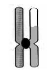
Meiosis is a type of cell division whereby a diploid cell (somatic cell) undergoes two cell divisions, and divides to form four dissimilar haploid cells (sex cells). Diploid cells have two sets of chromosomes, where each chromosome has a homologous partner. Haploid cells only have one set of chromosomes. Chromosomes in haploid cells have no homologous
partners.
Before meiosis begins (during interphase), DNA replication takes place. The result is two sets of chromosomes consisting of two identical chromatids joined together with a centromere. This is shown in Figure 2.1 (below).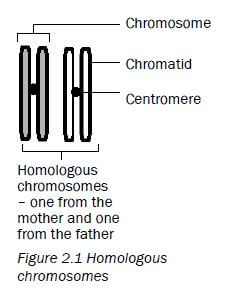
2.2 The process of meiosis in animal cells
Meiosis is the type of cell division used to produce gametes or sex cells (sperm and egg cells). A cell undergoing meiosis will divide twice - the first division is meiosis I and the second is meiosis II. | 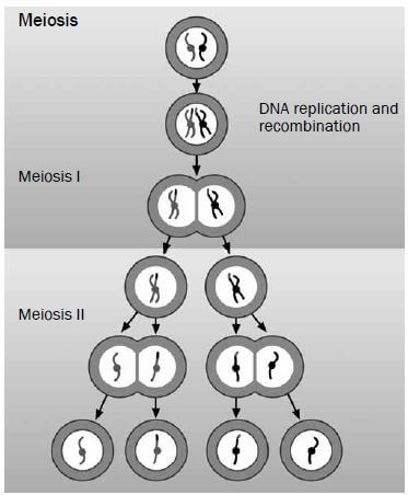 |
2.2.1 First meiotic division
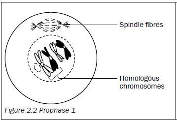
| Prophase 1
|
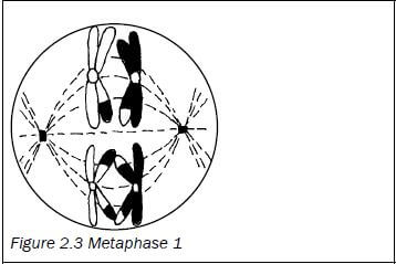
| Metaphase 1
|
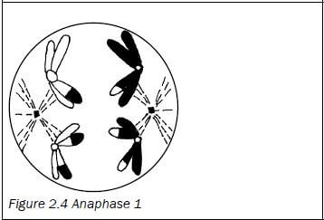
| Anaphase 1
|
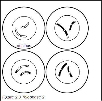
| Telophase 1
|

2.2.2 Second meiotic division
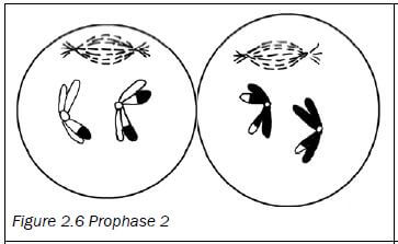
| Prophase 2
|
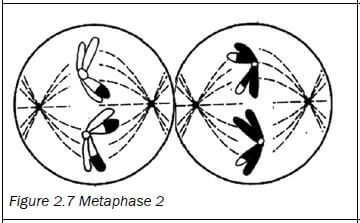
| Metaphase 2
|
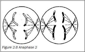
| Anaphase 2
|
| Telophase 2
|
An easy way to remember the events of meiosis is to use the word mnemonic IPMAT.
Letter | Phase | Event |
I | Interphase | I for in between: The part of the life cycle of the cell that is in between cell divisions. |
P | Prophase | P for preparation: The chromosomes prepare for meiosis by untangling and becoming clearly visible. Crossing over also takes place. |
M | Metaphase | M for middle: The chromosomes move to the ‘middle’ (equator). |
A | Anaphase | A for apart: The chromosomes/chromatids move apart/move to the poles. |
T | Telophase | T for terminal: The final phase of meiosis I/ meiosis II. |
2.3 The significance of meiosis
There are two reasons why meiosis is important.
- It reduces the number of chromosomes by half, in other words from diploid to haploid. This ensures that sex cells have half the number of chromosomes of other somatic cells so that when fertilisation occurs the zygote formed has the correct number of chromosomes. It balances the doubling effect of fertilisation.
- Crossing over introduces genetic variation. Genetic variation results in offspring that are better adapted to a particular environment and ensures that they will have a better chance of survival.
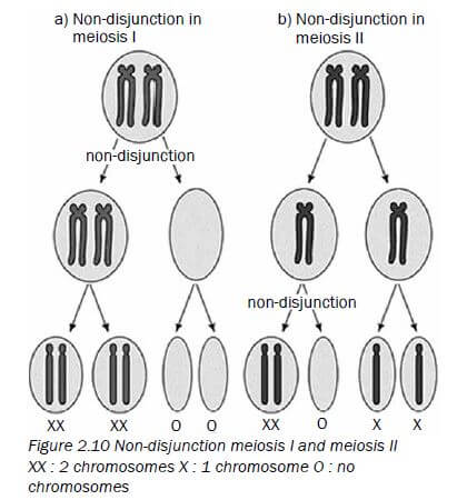
2.4 Abnormal meiosis
- Sometimes mistakes occur during the process of meiosis.
- This can happen in Anaphase 1 where the homologous chromosomes may not separate. Also called non-disjunction. (see above)
- It can also happen in Anaphase 2 when there is non-disjunction of the sister chromatids.
- If there is non-disjunction of chromosome pair 21 in humans it leads to the formation of an abnormal gamete with an extra copy of chromosome 21.
- If there is fusion between a normal gamete and an abnormal gamete (with extra copy of chromosome 21) it leads to Down Syndrome.
2.5 Differences between meiosis I and meiosis II
Meiosis I | Meiosis II |
The chromosomes arrange at the equator of the cell in homologous pairs. | Chromosomes line up at the equator of the cell individually. |
Whole chromosomes move to opposite poles of the cell. | Daughter chromosomes/chromatids move to opposite poles of the cell. |
Two cells form at the end of this division. | Four cells are formed at the end of this division. |
The chromosome number is halved during meiosis I. | The chromosome number remains the same during meiosis II. |
Crossing over takes place. | Crossing over does not take place. |
Table 2.1 The differences between meiosis I and meiosis II
Worked example
Study the diagrams below of two stages of meiosis then answer the questions that follow.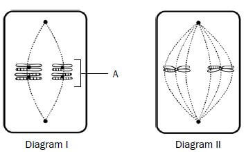
Figure 2.11 Two stages of meiosis
- State ONE visible reason in Diagram I which indicates that meiosis is taking place. (1)
- How many chromosomes would be present in each daughter cell at the end of meiosis in this cell? (1)
- Describe what takes place in the cell after the phase shown in Diagram I. (3)
- Tabulate TWO visible differences between the phases of meiosis shown in Diagrams I and II. (5) [10]
Answers to worked example
- The chromosomes are lined up at the equator of the cell in their homologous pairs.✔
OR
The chromosomes show evidence of crossing over.✔ (1) - Two ✔ chromosomes. (1)
- The next phase is Anaphase 1. The spindle fibres contract.✔ (shorten) and pull each chromosome ✔ of each chromosome pair to opposite poles ✔ of the cell.
(5) [10]Diagram I (metaphase 1)
Diagram II (metaphase 2)
1. Chromosomes are lined up at the equator in homologous pairs.✔
1. Chromosomes are lined up at the equator individually.✔
2. Four chromosomes are present✔
2. Two chromosomes are present✔
Activity 1
Question 1
Give the correct word or term for each of the statements or definitions provided below.
1.1 | The structure that joins the two halves of a double-stranded chromosome (1) |
1.2 | A pair of chromosomes, one inherited from each parent, that have the same genes at the same locus (1) |
1.3 | A single-stranded chromosome formed during Anaphase 2 (1) |
1.4 | The point of contact between two chromosomes of a homologous pair during crossing over (1) |
1.5 | One half of a double-stranded chromosome (1) |
1.6 | The phase in meiosis where crossing over occurs (1) [6] |
Answers to question 1
1.1 Centromere✔(1)
1.2 Homologous chromosomes✔ (1)
1.3 Daughter chromosome/chromatid✔ (1)
1.4 Chiasma✔/chiasmata✔ (1)
1.5 Chromatid✔ (1)
1.6 Prophase 1✔ (1) [6]
Question 2
Figure 2.12 (right) represents a process taking place during meiosis. Study the diagram and answer the questions that follow.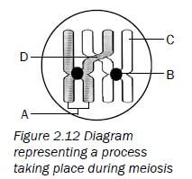
2.1 Provide labels for parts A, B, C and D. (4)
2.2 Name the process in meiosis that is illustrated in Figure 2.12. (1)
2.3 State ONE importance of the process you named in question 2.2. (2)
2.4 Draw a diagram of the structure labelled A to show its appearance immediately after the process you named in question 2.2. (2) [9]
Answers to question 2
2.1
A - Chromosome✔
B - Centromere✔
C - Chromatid✔
D - Chiasma✔/chiasmata (4)
2.2 Crossing over✔ (1)
2.3 It introduces genetic✔ variation✔ (2)
2.4
- A double-stranded chromosome with the strands joined by a centromere✔
- There is evidence of crossing over.✔ (2)

Question 3
Figure 2.13 (below) represents an animal cell in a phase of meiosis. Study the diagram and answer the questions that follow.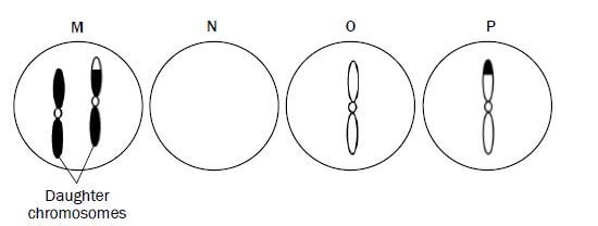
3.1 State whether the phase of meiosis shown in Figure 2.13 is meiosis I or meiosis II. (1)
3.2 Give ONE visible reason for your answer in question 3.1. (1)
3.3 Identify the parts labelled A and B. (2)
3.4 How many chromosomes:
- were present in the parent cell before meiosis began? (1)
- will be present in each cell at the end of meiosis? (1)
3.5 State ONE place in a human female where meiosis would take place. (1)
3.6 Could the cell represented in Figure 2.13 be that of a human? (1)
3.7 Explain your answer to question 3.6. (2)
3.8 Give TWO reasons why meiosis is biologically important. (2)
3.9 Give the term for the situation when some of the chromosomes do not separate correctly during the phase shown in Figure 2.13. (1) [13]
Answers to question 3
3.1 Meiosis II✔ (1)
3.2 Daughter chromosomes/chromatids are being pulled to the opposite poles✔ (1)
3.3
A - Spindle fibre✔
B - Cell membrane✔ (2)
3.4
a) 8✔
b) 4✔ (2)
3.5 Ovaries✔ (1)
3.6 No✔ (1)
3.7 There are only 4 chromosomes present3 instead of 23.✔ (2)
3.8
- It introduces genetic variation.✔
- It balances the doubling effect of fertilisation as it halves the number of chromosomes in the sex cells.✔ (2)
3.9 Non-disjunction✔ (1) [13]
Question 4
The diagram below shows the nuclei of the four cells that resulted from meiosis of chromosome pair 21 in a woman.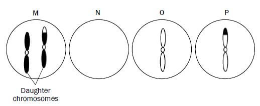
Figure 2.14: Diagram that shows the nuclei of four cells resulted from meiosis
4.1 Explain why nucleus N does NOT have a chromosome pair 21. (2)
4.2 Name and explain the disorder that will result if diagram M represents an egg cell that fuses with a normal sperm cell. (3) [5]
Answers to question 4
4.1 During Anaphase 1 the chromosome pair 21 does not separate✔/ non-disjunction. Gamete M will have an extra copy of chromosome number 21 and therefore gamete N does not have a copy of chromosome 21✔ (2)
4.2 Down syndrome✔/Trisomy 21 if gamete M fuses with normal sperm having 1 copy of chromosome 21 ✔ the resulting zygote will have 3 copies of chromosome 21✔ (3) [5]
For more papers on Meiosis visit https://www.elimuza.com/