Biological Concepts - Agricultural Sciences Grade 10 Study Guide
Share via Whatsapp Join our WhatsApp Group Join our Telegram GroupUNITS
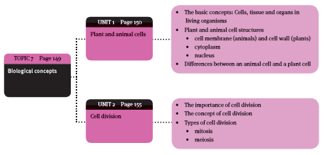
TOPIC 7: BIOLOGICAL CONCEPTS
Overview
What you will cover in Topic 7
Unit 1: Plant and Animal Cells
1.1 Basic concepts: cells, tissue and organs in living organisms
1.1.1 Cells
All living organisms are made up microscopic cells.
- Unicellular = consists of one cell (amoeba).
- Multicellular = consists of millions – billions of cells (humans)
- All cells have a cell membrane.
- Inside the cell is a jelly-like fluid = cytoplasm.
- Organelles are suspended in the cytoplasm.
- Each organelle has a special function in the cell.
1.1.2 Tissue
A tissue is a group of cells that have the same structure and function.
- Animals are composed of tissue, such as nerve tissue and muscle tissue. Animals also contain blood, which is the only liquid tissue in animals.
- Plants contain different kinds of tissue, such as parenchyma and cork.
1.1.3 Organs
An organ = a group of tissues that have the same structure and function.
- Animals have organs, e.g. a heart, a brain, a stomach and a liver.
- Plants have organs, e.g. roots, stems, leaves and fruit.
1.1.4 Organisational levels of multicellular organisms
The relationship between cells, tissue and organs show how a multicellular organism is organised.
- The first level of organisation is the cell.
- The highest level of organisation is the organ.
1.2 Plant and animal cell structures
Plant and animal cells are alike in many ways, but there are some differences.
- Animal cells differ from one another, according to their own specialised function, for example, cells in liver tissue are different from cells in lung tissue.
- Plant cells differ from one another according to their function in the leaf, stem, root or flower.
- Cells also differ in size: from 2–15 micrometres in diameter (micrometre / micron, is one thousand times smaller than a mm: symbol = μm.)
Cells have three main parts or components:
- cell membrane (animals cells) / cell wall (plant cells)
- cytoplasm
- nucleus.
1.2.1 Cell membrane (animal cells)
- Also called the plasmalemma or plasma membrane.
- made up of protein and fats.
- very thin, with tiny pores that allow certain substances to go into or out of the cell = semi-permeable.
1.2.2 Cell wall (plant cells)
- Only plant cells have a cell wall = one of the main differences between plant and animal cells.
- The cell wall is situated outside the cell membrane.
- The cell wall is not living tissue.
- It is made up of cellulose.
- Each plant cell has a primary cell wall, but as the cell ages, each plant cell forms secondary and tertiary cell walls:
- older cells have thicker walls than younger cells
- with woody plants, the outer two cell wall layers contain lignin, which makes the cell wall rigid and solid.
- When secondary and tertiary cell walls form, no extra cellulose is deposited in certain places → known as pits.
- The pits allow cytoplasmatic threads (plasmodesmata) to go through from one plant cell to the next. The cytoplasmatic threads (plasmodesmata) allow water, glucose and amino acids to pass from one cell to the other.
- Two plant cells are separated by another type of wall = middle lamella → made up of pectose.
1.2.3 Cytoplasm
- Cytoplasm is the jelly-like fluid content in the cell.
- The cytoplasm contains organelles: each has its own function.
1.2.4 The nucleus
The nucleus is the ‘control centre’ of a cell. It is situated in the centre of the cell.
- The nucleus is surrounded by a double membrane and nucleoplasma (fluid).
- The nucleus contains nucleoli. Each nucleolus contains RNA molecules that help with the production of proteins.
- The nucleoplasma contains the chromatin network, which contains the DNA of the cell. DNA contains the hereditary material of the cell and determines what the cell looks like. The chromatin network plays a very important role during cell division.
1.2.5 Cell organelles and their main functions
The cytoplasm of animal and plant cells has various organelles suspended in them:
- Endoplasmic reticulum (ER)
- Ribosomes
- Mitochondrion
- Golgi apparatus
- Lysosomes
- Plastids
- Vacuoles
Endoplasmic reticulum (ER)
Endoplasmic reticulum (ER) = a network of fine membranes that look like tubes. These tubes connect the cell membrane with the outer membrane of the nucleus.
- The ER is a transport system inside a cell.
- Different substances move through the ER inside of the cell.
- Sometimes there are small particles on the surface of the ER → called ribosomes.
- ER with ribosomes is called ribosomal ER or ‘rough ER’.
- Sometimes there are no ribosomes on the ER.
- The ER is then called ‘smooth ER’.
Ribosomes
- Ribosomes are found on the surface of the ER.
- Some ribosomes can occur in small groups in the cytoplasm.
- Such ribosomes are called polyribosomes (‘poly’ = many).
- They consist of RNA and contain enzymes.
- They use the enzymes to make proteins out of amino acids.
- The mitochondrion is the ‘powerhouse’ of a cell.
- The process of respiration takes place inside the mitochondrion.
- During this process, molecules that contain energy are broken down and the energy is released.
- The cell can use the energy to perform different functions.
- Cells that need more energy (e.g. cells is muscle tissue) have much more mitochondria than cells that do not need much energy.
- Mitochondria have an oval or sausage shape.
- Their walls consist of two membranes:
- the inner membrane and the outer membrane.
- The inner membrane has folds → called cristae.
- Respiration takes place on the surface of the cristae.
- The space between the two membranes is filled with liquid.
- the inner membrane and the outer membrane.
Golgi apparatus
Golgi apparatus = a collection of membranes stacked on top of one another (like pancakes).
- Golgi apparatus is normally situated close to the nucleus and works closely with the endoplasmic reticulum.
- The Golgi apparatus modifies proteins and brings them to the cell surface where they can be secreted.
- Secretions include hormones, enzymes, antibodies and other molecules.
- Cells that have a secretory function, for example gland cells, contain more of these organelles than other cells.
Lysosomes
- Similar to mitochondria.
- Sac-like structures that have two membranes.
- More often found in animal cells than plant cells.
- Liver cells contain many lysosomes.
- Lysosomes contain various enzymes that help with the digestion of large molecules, such as protein.
- These enzymes are responsible for the destruction of old cells.
- This is known as autolysis.
Plastids
- Only appear in plant cells.
- They vary in size and can be round, oval or disc-shaped.
- There are three types of plastids:
- Chloroplasts: in the cells of all the green parts of a plant → contain chlorophyll, which gives plants their green colour. Chloroplasts are oval in shape and are surrounded by a single membrane. They also have an inner membrane. Photosynthesis takes place in the chloroplasts.
- Chromoplasts: Chromoplasts are responsible for the red, orange and yellow colour of many flowers, fruits and autumn leaves. Often called the ‘colour plastids’. The colour is produced by a number of pigments. The most important pigments = carotenoids and xantophylls. Chromoplasts are important because:
- bright colours of flowers attract insects for pollination
- bright colours of fruits attract insects and animals that help in the distribution of seeds
- animals produce vitamin A from the carotenoids that they eat.
- Leucoplasts: plastids that contain no colour or chlorophyll. They only appear in cells that are not exposed to sunlight (e.g. the cells in the roots and seeds of plants). If leucoplasts are exposed to sunlight, they change into chloroplasts and start to produce chlorophyll (e.g. potatoes that start to turn green if they are exposed to sunlight). The main functions of leucoplasts = store nutrients (such as starch) and to manufacture oils and some proteins.
Vacuoles
Vacuoles are hollow spaces in the cytoplasm filled with a watery fluid called cell sap.
- Occur in plant and animal cells, but are more well-developed in plant cells.
- Older cells have larger vacuoles than younger cells. Eventually, the vacuoles join to form one large, central vacuole.
- The tonoplast, a cytoplasmic membrane, surrounds the vacuoles. The cell sap consists of water, in which substances like sugars, salts, pigments and gases are dissolved.
1.3 Differences between a plant cell and an animal cell
Difference between plant and animal cells | ||
Factor or part | Plant cell | Animal cell |
General appearance | Large, with distinct boundaries | Smaller, with indistinct boundaries |
Cell boundary | Distinct cell wall of cellulose present on both sides of the middle lamella | No cell wall; only a living cell membrane |
Space between cells | Usually present | None |
Lysosomes | None | Contains lysosomes, especially liver cells |
Plastids | Occur commonly | None |
Vacuoles | Common in plant cells. Occupies the largest part of the cell | Much smaller, not always clear |
Starch grains | Usually occur | None; contains oil globules |
Protein | Usually none | Usually occurs |
The differences between an animal cell and a plant cell
Unit 2: Cell Division
2.1 The importance of cell division
Cell division is perhaps the most important process in living cells.
- All living creatures (unicellular or multicellular) begin life as one cell (zygote).
- Almost immediately, this cell divides to form two cells (multicellular).
- The two cells divide to form four cells, and so on.
- Cell division takes place throughout the life of any living organism to:
- repair damaged cells
- grow
- reproduce.
The study of cell division is very important:
- helps to find a cure for diseases such as cancer, because cancer is the uncontrollable division of cells.
- has enabled scientists to perform artificial insemination of cattle, as well as cloning.
2.2 The concept of cell division
During cell division, a cell divides to form two cells. This means that information about the characteristics of the ‘mother cell’ must be passed on to the two ‘daughter cells’.
- Hereditary information is contained in the DNA (deoxyribonucleic acid) of the cell.
- The DNA and proteins are the chromatin network of the cell.
- When cell division is about to begin, the chromatin network forms tightly coiled strands, called chromatids.
- Two such chromatids are joined by means of a centromere to form a chromosome.
- Chromosomes are a very important part of cell division.
- The cytoplasm and cell membrane of the cell must also divide. This happens during the process of cytokinesis.
2.3 Types of cell division
2.3.1 Mitosis (in all body cells)
Each living cell grows until it reaches its maximum size. Then the adult cell (or ‘mother cell’) divides to form two new cells (or ‘daughter cells’). In this way, new cells are formed that are necessary for growth and for replacing old cells.
There are four phases to mitosis:
- prophase
- metaphase
- anaphase
- telophase.
The prophase (first phase)
- The cell membrane starts to break down
- The chromatin network starts changing. The threads become shorter and thicker and are now called chromatids.
- Two chromatids are joined by a centromere to form a chromosome.
- Each different species has a certain number of chromosomes.
- The human body has 46 chromosomes in each cell.
- Each different species has a certain number of chromosomes.
- The nuclear sap changes from a fluid to a gel
- Larger cell structures, such as the plastids (plant cells) and mitochondria disappear.
- The nucleolus disappears.
- Spindle threads start forming in the cytoplasm.
Metaphase (middle phase)
- First, the chromosomes start arranging themselves so that the centromeres are along the middle of the cell (or ‘equator’)
- Then, some spindle threads stretch from one end (or ‘pole’) of the cell to the other (continuous spindle threads).
- Other spindle threads are attached to the chromatids and are called chromosomal spindle threads.’
Anaphase (separation phase)
- The centromere of each chromosome divides into two equal parts.
- The two chromatids of each chromosome start moving away from each other towards the opposite poles, pulled along by the chromosomal spindle threads.
- As soon as the chromatids start to divide, they are known as daughter chromosomes.
Telophase (two new nuclei form)
- The spindle threads disappear.
- A nuclear membrane begins to develop around the chromosomes at each pole
- The chromosomes become thinner and lengthen → become the chromatin network of each of the two new cells
- A nucleolus appears in each nucleus
- In plant cells, the cell wall starts developing at the equator. In animal cells, a constriction starts → will eventually lead to the separation of the daughter cells
- At the end of this phase, the mother cell has divided into two identical halves. The chromosomes of the new daughter cells are exactly the same as those of the mother cell from which they formed.
- The cells have now reached the interphase.
- Between two episodes of mitosis, the cell rests and prepares for the next episode of mitosis. Mitoses and the interphase make up the cell cycle.
The importance of mitosis
- The zygote cannot grow without the process of mitosis.
- New cells must continuously be made to replace old cells.
- This cannot take place without mitosis.
- Mitosis ensures that the daughter cells are identical to the mother cell.
- During mitosis chromosomes are duplicated, so the genetic material of the mother cell is also duplicated.
- The two daughter cells are identical to each other, but also to the mother cell from which they developed.
2.3.2 Meiosis
- Each species has a definite, specific number of chromosomes, for example:
- Humans have 46 chromosomes (23 pairs) in each cell
- Horses have 64 chromosomes (32 pairs) in each cell
- Sheep have 54 chromosomes (27 pairs) in each cell.
- Meiosis takes place in sex cells (also called gametes), and not in other body cells.
- During meiosis, the number of chromosomes is halved.
- So each male sex cell (sperm cell) and female sex cell (ovum or egg cell) only contains 23 chromosomes each.
- When a sperm cell fertilises an egg cell, the zygote that forms has 46 chromosomes.
- If the number of chromosomes in a sex cell were not halved during meiosis, the zygote would have 92 chromosomes, instead of 46 chromosomes.
- Therefore, during meiosis, a diploid set of chromosomes (2n) is reduced to a haploid set of chromosomes (n).
- This ensures that the zygote that is formed as a result of fertilisation has a diploid set of chromosomes → keeps the number of chromosomes the same from one generation to the next.
Meiosis occurs in two stages that follow one another in rapid succession.
First meiotic division
The phases of division are described by the same names as with mitosis.
Prophase
The prophase of meiosis is more complicated than that of mitosis. With meiosis:
- The nuclear membrane and the nucleolus disappear.
- Spindle threads form and meet at the poles of the cell.
- The chromatin network shrinks and the chromosomes become visible as separate threads.
- The chromosomes synapse (or combine) in homologous pairs.
- Homologous pairs are pairs of the same chromosome.
- Each chromosome then forms a bivalent (also known as a tetrad).
- A bivalent is a homologous pair of chromosomes that is wound around each other. In other words, a bivalent is two chromosomes with two centromeres.
- Homologous pairs are pairs of the same chromosome.
- The chromosomes then shrink and become thicker.
- Each chromosome of the bivalent then unravels lengthwise to become two chromatids, so that each bivalent contains four chromatids. The centromeres do not divide.
- Now a crossing over process takes place between chromatids at points called chiasmata. Chromatids break and rejoin, so that each chromatid joins to its homologous partner. Crossing over allows the chromosomes to exchange genetic material, allowing for more different combinations of genetic material.
Metaphase
- The bivalents arrange themselves on the equator of the cell.
- The two centromeres of each bivalent are on either side of the equator.
Anaphase
The two chromosomes of each bivalent move to opposite poles.
Telophase
- First, the chromosomes regroup at the poles. Each of the chromosomes consists of two chromatids joined together by a centromere.
- Next, two separate cells develop, each with only half the number of chromosomes of the parent nucleus.
Second meiotic division
- The second meiotic division happens just after the first meiotic division (separating the two chromatids of each chromosomes)
- The second meiotic division is usually the same as ordinary mitosis, except that the prophase is of very short duration.
- Therefore, the second meiotic division actually only really starts with metaphase.
Prophase II
- No significant changes take place in the chromosomes.
- The DNA in the cell does not replicate.
- Nuclear membrane and nucleolus disappear.
- Asters and spindle fibres are formed.
Metaphase II
The chromosomes arrange themselves along the equator of the cell, forming a single metaphase plate (as in mitosis).
Anaphase II
- Each centromere divides.
- The two daughter chromatids of each chromosome move to opposite poles of the cell.
- Finally, they reach the poles of the cell. Each pole now has haploid number of chromosomes and half the amount of DNA.
Telophase
- At the poles, the daughter chromosomes form a new chromatin network.
- The nuclear membrane and nucleolus reappear.
- In plant cells, a cell plate is formed along the equator. In animal cells, a constriction appears.
- The mother cell has now divided into four cells that are all different from the mother cells and from each other.
The importance of meiosis
- Meiosis reduces the diploid number of chromosomes to a haploid number. This ensures that the number of chromosomes of an organism stays constant from one generation to the next.
- Meiosis makes the exchange of chromatin segments possible, thereby mixing genes. If this did not happen, parents and children – as well as siblings in a family – would have been almost like clones of one another.
2.4 The differences between mitosis and meiosis
The table below shows the differences between mitosis and meiosis.
Differences between mitosis and meiosis | |
Mitosis | Meiosis |
Takes place in somatic cells (cells in the body) | Takes place in the sex cells (gametes) |
One division of the mother cell gives two daughter cells, identical to one another and the mother cell | Two divisions of the mother cell give four haploid gametes, each different from one another and the mother cell |
Number of chromosomes in the nucleus of mother and daughter cells remain the same | Mother cell has a diploid (2n) number of chromosomes. The four gametes are all haploid (n). Therefore, the number of chromosomes in the mother cell is halved. |
No pairing of homologous chromosomes | During prophase of the first meiotic division, pair- ing of homologous chromosomes takes place |
No crossing-over between chromosomes | Crossing-over takes place |
Centromeres split during anaphase | Centromeres only split during anaphase of the second meiotic division (and not during the anaphase of the first meiotic division) |
The daughter cells have exactly the same genetic make up as the mother cell | The gametes have different genetic material than the mother cell |
The differences between mitosis and meiosis
Topic 7: Questions
- Answer the questions below.
- Give yourself one hour.
- Check your answers afterwards and do corrections.
Questions
- Draw a diagram of a plant cell with its main components. (8)
- What are the following called? (5)
- Cytoplasmic threads in plant cells.
- The separating wall between two plant cells.
- The jelly-like fluid content in the cell.
- Ribosomes that occur in small groups in the cytoplasm.
- The watery fluid in vacuoles.
- State where plastids occur, give a brief description of the three types and state the function of each. (9)
- What is the function of the cytoplasmic threads? (3)
- Draw a diagram of a plant cell and name all the parts. (20)
- What are the three main functions of cell division in a living organism? (3)
- Describe the process that takes place during the prophase of mitosis. (8)
- What are the differences between meiosis and mitosis? (14)
[Total marks: 70]